. 2010 May;16(5):580-5, 1p following 585.
doi: 10.1038/nm.2122. Epub 2010 Apr 25.

PU.1-mediated upregulation of CSF1R is crucial for leukemia stem cell potential induced by MOZ-TIF2

Affiliations

PU.1-mediated upregulation of CSF1R is crucial for leukemia stem cell potential induced by MOZ-TIF2

Yukiko Aikawa et al. Nat Med. 2010 May.

Abstract

Leukemias and other cancers possess self-renewing stem cells that help to maintain the cancer. Cancer stem cell eradication is thought to be crucial for successful anticancer therapy. Using an acute myeloid leukemia (AML) model induced by the leukemia-associated monocytic leukemia zinc finger (MOZ)-TIF2 fusion protein, we show here that AML can be cured by the ablation of leukemia stem cells. The MOZ fusion proteins MOZ-TIF2 and MOZ-CBP interacted with the transcription factor PU.1 to stimulate the expression of macrophage colony-stimulating factor receptor (CSF1R, also known as M-CSFR, c-FMS or CD115). Studies using PU.1-deficient mice showed that PU.1 is essential for the ability of MOZ-TIF2 to establish and maintain AML stem cells. Cells expressing high amounts of CSF1R (CSF1R(high) cells), but not those expressing low amounts of CSF1R (CSF1R(low) cells), showed potent leukemia-initiating activity. Using transgenic mice expressing a drug-inducible suicide gene controlled by the CSF1R promoter, we cured AML by ablation of CSF1R(high) cells. Moreover, induction of AML was suppressed in CSF1R-deficient mice and CSF1R inhibitors slowed the progression of MOZ-TIF2-induced leukemia. Thus, in this subtype of AML, leukemia stem cells are contained within the CSF1R(high) cell population, and we suggest that targeting of PU.1-mediated upregulation of CSF1R expression might be a useful therapeutic approach.


Figures
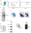
Fig. 1
Fig. 1. CSF1Rhigh cells show potent leukemia-initiating activity
(a) The BM cells from MOZ-TIF2–induced AML mice were analyzed by FACS for expression of GFP and CSF1R. (b) CSF1Rhigh and CSF1Rlow/− cells were sorted by flow cytometry, and MOZ-TIF2 expression was investigated by immunoblot analysis using an anti-MOZ antibody. (c, d) The indicated numbers of flow-sorted CSF1Rhigh (c) and CSF1Rlow/− (c) cells were transplanted into sub-lethally irradiated mice, and leukemia-free survival was investigated. n = 6, P = 0.0001 (104, 103 and 102) and 0.3173 (101). Three independent experiments were performed and essentially the same results were obtained. (e) The BM cells from MOZ-TIF2–induced AML mice were analyzed by FACS for expression of Mac1 and CSF1R. (f–h) CSF1Rhigh and CSF1Rlow/− cells were sorted and analyzed for morphology by staining with May-Giemsa (f), colony-forming activity in methylcellulose medium (g), and levels of total and phosphorylated STAT5, phosphorylated ERK, and PU.1 (h). The scale bars represent 10 ?m in (f). (i) The BM cells from an AML patient with t(8;16) were cultured for 3 d in 10 ng/ml hM-CSF and tested by FACS for CSF1R expression. (j) CSF1Rhigh and CSF1Rlow/− cells of the AML patient with t(8;16) were sorted and analyzed for MOZ-CBP transcripts by RT-PCR.
Fig. 2
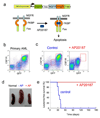
Fig. 2. Cure of AML by ablation of CSF1Rhigh cells
(a) Structure of genes for the CSF1R promoter, EGFP, the NGFR–FKBP–Fas suicide construct, and activation of NGFR–FKBP–Fas. In transgenic (Csf1r-EGFP-NGFR/FKBP1A/TNFRSF6) mice, conditional ablation of cells expressing high levels of CSF1R can be induced by exposure to the AP20187 dimerizer. (b) The BM cells from transgenic mice were infected with MSCV-MOZ-TIF2-ires-GFP and were transplanted into lethally irradiated C57BL/6 mice to induce AML. Expression levels of GFP and CSF1R in BM cells were analyzed by flow cytometry two months after transplantation. (c–e) Bone marrow cells (1 × 10⁵) of primary AML mice were transplanted into sub-lethally irradiated C57BL/6 mice. Administration of AP20187 or solvent (control) to the secondary AML mice was started by IV injection three weeks after transplantation. Expression of GFP and CSF1R in BM cells (c) and spleen sizes (d) were analyzed four weeks after transplantation. Bars represent 1 cm. Leukemia-free survivals of the untreated (n = 6) and AP20187-treated (n = 6) secondary transplanted mice were also investigated (e). P < 0.0001
Fig. 3
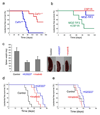
Fig. 3. Roles of CSF1R in MOZ-TIF2–induced AML
(a) Fetal liver cells of E16.5 CSF1R+/+ and CSF1R−/− mice embryo littermates were infected with either MOZ-TIF2-ires-GFP, and transplanted into irradiated mice. The leukemia-free survivals of the mice were analyzed. n = 8, P < 0.0001 (b) Fetal liver cells of CSF1R+/+ mice were infected with CSF1R, MOZ-TIF2, or both, and transplanted into irradiated mice. n = 5 (c–e) The BM cells (105) from AML mice with MOZ-TIF2 (c, d) or N-MYC (e) were transplanted into irradiated mice. Imatinib mesylate was administrated twice daily. Spleen sizes of the MOZ-TIF2–introduced mice were analyzed three weeks after transplantation (C). Bars represent 1 cm. Leukemia-free survivals of the mice were analyzed (d, e). (d) n = 8, P < 0.0001 (+Ki20227 and +Imatinib) (e) n = 8, P = 0.3825 (+Ki20227) and 0.4051 (+Imatinib)
Fig. 4
Fig. 4. PU.1-dependent upregulation of CSF1R by MOZ-fusions
(a) Protein-interacting domains on MOZ and PU.1. (b) Effects of MOZ, MOZ-CBP, and MOZ-TIF2 on AML1- and PU.1-mediated transcription of the CSF1R promoter. SaOS2 cells were transfected with the CSF1R–luciferase construct and the effectors indicated. Luciferase activity was analyzed 24 h after transfection. Error bars represent SD (n = 3). * P < 0.01 and ** P < 0.005. Six independent experiments were performed and essentially the same results were obtained. (c) SaOS2 cells were transfected with the wild-type CSF1R–luciferase construct or its mutant lacking the PU.1-binding site, together with effectors indicated. (d) PUER cells infected with MSCV-GFP or MSCV-MOZ-TIF2-ires-GFP retroviruses were exposed to 100 nM 4-hydroxytamoxifen (4-HT) for 0, 2, or 5 d and analyzed by FACS for CSF1R expression. Three independent experiments showed the same results. (e–g) Fetal liver cells of E12.5 PU.1+/+ and PU.1−/− mouse embryo littermates were infected with either MOZ-TIF2 (e) or N-MYC (f), and transplanted into irradiated mice. Fetal liver cells of PU.1−/− mice were infected with PU.1, MOZ-TIF2, or both, and transplanted into irradiated mice (g). Leukemia-free survivals of the mice were analyzed. (e) n = 8, P < 0.0001 (F) n = 4, P = 0.0943 (g) n = 5, P = 0.0001 (PU.1 + MOZ-TIF2 vs. others) (h) The fetal liver cells of E14.5 PU.1flox/flox with ER-Cre were infected with MOZ-TIF2, and transplanted into irradiated mice. The BM cells of the primary AML mice were transplanted into sub-lethally irradiated wild-type mice. Tamoxifene or solvent (control) was administered to the secondary AML mice every 2 d by intravenous injection 17 d after transplantation, when GFP+ cells were detected in peripheral blood. Leukemia-free survivals of the secondary mice were investigated. n = 5, P = 0.0018 (i) Model for transcriptional regulation by normal and fusion MOZ proteins. MOZ-fusions stimulated constitutive CSF1R expression to induce leukemia (left panel). Normal MOZ controlled CSF1R expression by binding to PU.1 to regulate normal hematopoiesis (right panel).

References

    1. Bonnet D, Dick JE. Human acute myeloid leukemia is organized as a hierarchy that originates from a primitive hematopoietic cell. Nat Med. 1997;3:730–737. - PubMed
    1. Reya T, Morrison SJ, Clarke MF, Weissman IL. Stem cells, cancer, and cancer stem cells. Nature. 2001;414:105–111. - PubMed
    1. Borrow J, et al. The translocation t(8;16)(p11;p13) of acute myeloid leukaemia fuses a putative acetyltransferase to the CREB-binding protein. Nat Genet. 1996;14:33–41. - PubMed
    1. Katsumoto T, Yoshida N, Kitabayashi I. Roles of the histone acetyltransferase monocytic leukemia zinc finger protein in normal and malignant hematopoiesis. Cancer Sci. 2008;99:1523–1527. - PMC - PubMed
    1. Katsumoto T, et al. MOZ is essential for maintenance of hematopoietic stem cells. Genes Dev. 2006;20:1321–1330. - PMC - PubMed

Publication types

MeSH terms

Substances