Review
. 2010 May;7(5):277-90.
doi: 10.1038/nrcardio.2010.32.

Fetal cardiac arrhythmia detection and in utero therapy

Affiliations
Review

Fetal cardiac arrhythmia detection and in utero therapy

Janette F Strasburger et al. Nat Rev Cardiol. 2010 May.

Abstract

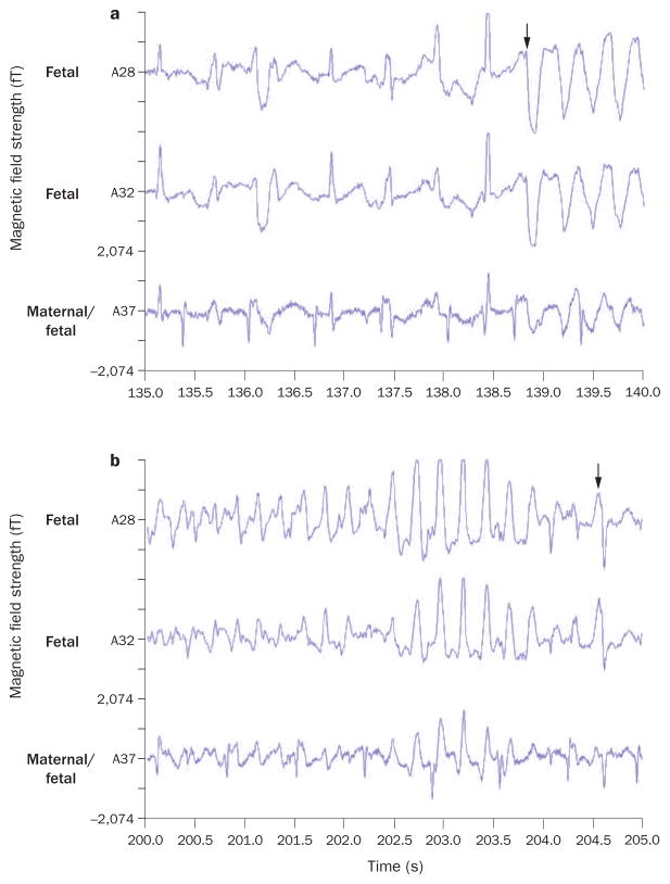
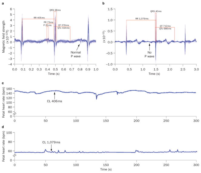
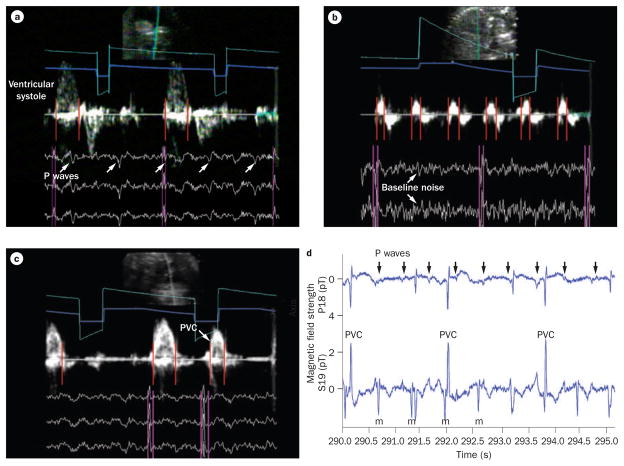
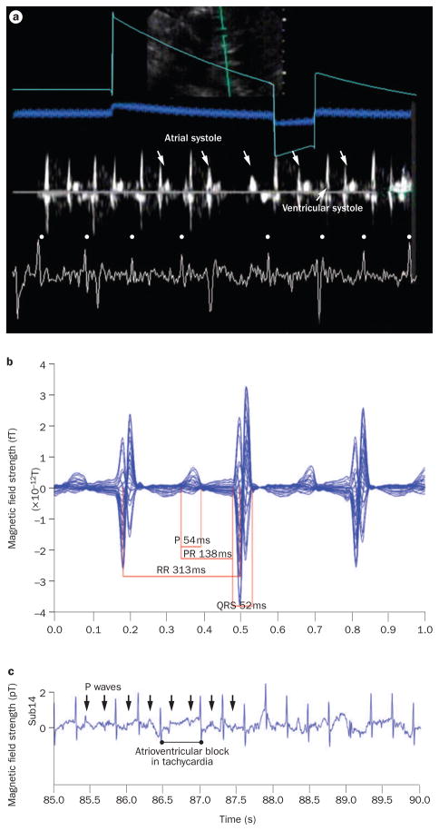
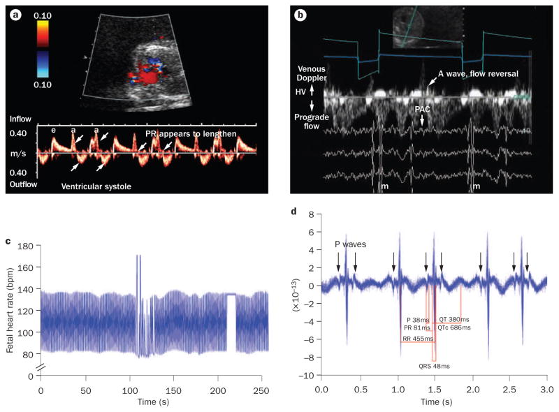
The human fetal heart develops arrhythmias and conduction disturbances in response to ischemia, inflammation, electrolyte disturbances, altered load states, structural defects, inherited genetic conditions, and many other causes. Yet sinus rhythm is present without altered rate or rhythm in some of the most serious electrophysiological diseases, which makes detection of diseases of the fetal conduction system challenging in the absence of magnetocardiographic or electrocardiographic recording techniques. Life-threatening changes in QRS or QT intervals can be completely unrecognized if heart rate is the only feature to be altered. For many fetal arrhythmias, echocardiography alone can assess important clinical parameters for diagnosis. Appropriate treatment of the fetus requires awareness of arrhythmia characteristics, mechanisms, and potential associations. Criteria to define fetal bradycardia specific to gestational age are now available and may allow detection of ion channelopathies, which are associated with fetal and neonatal bradycardia. Ectopic beats, once thought to be entirely benign, are now recognized to have important pathologic associations. Fetal tachyarrhythmias can now be defined precisely for mechanism-specific therapy and for subsequent monitoring of response. This article reviews the current and future diagnostic techniques and pharmacologic treatments for fetal arrhythmia.


Conflict of interest statement

Competing interests

The authors and the Journal Editor B. Mearns declare no competing interests. The CME questions author D. Lie has served as a nonproduct speaker for “Topics in Health” for Merck Speaker Services.

Figures

Figure 1
Figure 1
Two fetal magnetocardiography recordings from a 30-week fetus with long QT syndrome 3. a | Onset of torsades de pointes ventricular tachycardia occurs after 138.5 s (arrow). b | Termination of torsades de pointes occurs at 204.5 s (arrow). Arrhythmia was triggered by use of amiodarone to treat what was initially considered, on the basis of findings from echocardiography, to be drug-refractory fetal ventricular tachycardia. Permission obtained from Elsevier Ltd. © Cuneo, B. F. et al. Am. J. Cardiol. 91, 1395–1398 (2003).
Figure 2
Figure 2
Signal-averaged electrograms of fetal magnetocardiography recordings from a fetus with maternal SSA/Ro autoantibody isoimmunization. a | Sinus rhythm is present at 19 weeks of gestation. b | At 20 weeks, complete atrioventricular block had developed with a prolonged QTUc interval of 686 ms. The P wave appears absent because it is signal-averaged away when not stably associated with the QRS interval. c | At 19 weeks, the fetal heart rate trend over 5 min (arrow) documents stable sinus rhythm at ~145 bpm. d | At 20 weeks, the fetal heart rate trend over 5 min (arrow) shows third-degree atrioventricular block at 55 bpm. Abbreviation: CL, cycle length.
Figure 3
Figure 3
Simultaneous fetal magnetocardiography and Doppler echocardiography recordings from a fetus with isoimmune third-degree atrioventricular block at 23 weeks and 4 days of gestation. The two data types (echocardiographic and magnetocardiographic) were superimposed using timing marker channels (gray and turquoise lines) to provide a single image during postprocessing. QRS complexes (pink vertical lines) precede mechanical atrial wall and ventricular systolic velocities (red vertical lines), which enables interpretation of the electromechanics of the fetal heart. a | P waves are indicated by arrows and are not conducting. b | Baseline noise (arrows) results from Doppler echocardiography frequencies that must be removed during signal processing. Atrial wall velocities (red lines) and QRS complexes indicate atrioventricular dissociation. c | Ventricular systolic Doppler events, including a PVC (arrow). d | A fetal magnetocardiography recording from this fetus shows third-degree atrioventricular block and a narrow QRS duration with frequent ventricular ectopy. P waves (arrows) are not conducting. Abbreviations: m, maternal; PVC, premature ventricular contraction.
Figure 4
Figure 4
Combined fetal magnetocardiography and Doppler echocardiography tracing from a fetus with atrial ectopic tachycardia and intermittent atrioventricular block (during tachycardia) at 25 weeks and 5 days of gestation. a | A simultaneous echocardiography and fetal magnetocardiography tracing from this fetus showed atrial systole wall motion events (down-facing arrows) that documented the persistence of atrial activity during atrioventricular block of tachycardia. QRS complexes in the fetal magnetocardiography tracing are indicated by dots. b | Fetal magnetocardiography 20 s signal-averaged recording (1 s display). The atrial cycle length was ~313 ms (heart rate 192 bpm). c | A fetal magnetocardiography rhythm tracing from the same fetus, showing atrioventricular conduction block during tachycardia with perpetuation of atrial activity (arrows indicate P waves).
Figure 5
Figure 5
Fetal magnetocardiography and Doppler echocardiography tracings from a fetus with a rhythm pattern that simulated Mobitz type I second-degree atrioventricular block at 18 weeks and 1 day of gestation. This fetus presented with tachyarrhythmia (not shown) of 260 bpm. a | Doppler echocardiography of the left ventricular inflow and outflow shows that atrioventricular conduction appears prolonged and followed by dropped ventricular events (upward arrows) and hidden atrial events (downward arrows). b | Simultaneous fetal magnetocardiography and Doppler echocardiography demonstrates venous flow reversal (diagonal arrow) consistent with cannon A waves after alternate QRS complexes that are caused by blocked PACs (down-facing arrows). c | The fetal heart rate trends show persistence of this alternating rhythm over most of the 5 min recording. d | A 20 s signal-averaged trace of the QRS interval before the pause led to a final diagnosis of blocked atrial trigeminy. Abbreviations: a, atrial; e, passive inflow; HV, hepatic vein; PAC, premature atrial contraction.

References

    1. Cuneo BF, et al. Prenatal diagnosis and in utero treatment of torsades de pointes associated with congenital long QT syndrome. Am J Cardiol. 2003;91:1395–1398. - PubMed
    1. Hamada H, et al. Prenatal diagnosis of long QT syndrome using fetal magnetocardiography. Prenat Diagn. 1999;19:677–680. - PubMed
    1. Horigome H, Iwashita H, Yoshinaga M, Shimizu W. Magnetocardiographic demonstration of torsade de pointes in a fetus with congenital long QT syndrome. J Cardiovasc Electrophysiol. 2008;19:334–335. - PubMed
    1. Hosono T, Shinto M, Chiba Y, Kandori A, Tsukada K. Prenatal diagnosis of fetal complete atrioventricular block with QT prolongation and alternating ventricular pacemakers using multi-channel magnetocardiography and current-arrow maps. Fetal Diagn Ther. 2002;17:173–176. - PubMed
    1. Miller TE, et al. Recurrent third-trimester fetal loss and maternal mosaicism for long-QT syndrome. Circulation. 2004;109:3029–3034. - PubMed

Publication types

MeSH terms