. 2010 Apr 15;5(4):e10178.
doi: 10.1371/journal.pone.0010178.

T- and B-cell-mediated protection induced by novel, live attenuated pertussis vaccine in mice. Cross protection against parapertussis

Affiliations

T- and B-cell-mediated protection induced by novel, live attenuated pertussis vaccine in mice. Cross protection against parapertussis

Pascal Feunou Feunou et al. PLoS One. .

Abstract

Background: Despite the extensive use of efficacious vaccines, pertussis still ranks among the major causes of childhood mortality worldwide. Two types of pertussis vaccines are currently available, whole-cell, and the more recent acellular vaccines. Because of reduced reactogenicity and comparable efficacy acellular vaccines progressively replace whole-cell vaccines. However, both types require repeated administrations for optimal efficacy. We have recently developed a live attenuated vaccine candidate, named BPZE1, able to protect infant mice after a single nasal administration.

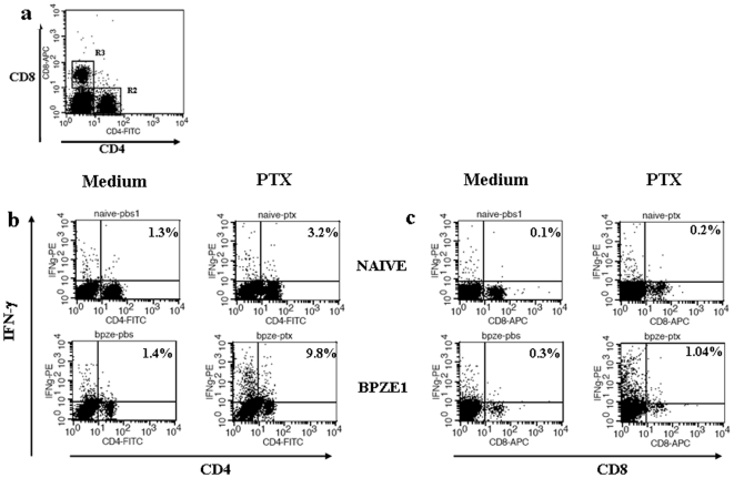
Methodology/principal findings: We determined the protective mechanism of BPZE1-mediated immunity by using passive transfer of T cells and antibodies from BPZE1-immunized mice to SCID mice. Clearance of Bordetella pertussis from the lungs was mediated by both BPZE1-induced antibodies and CD4(+), but not by CD8(+) T cells. The protective CD4(+) T cells comprised IFN-gamma-producing and IL-17-producing subsets, indicating that BPZE1 induces both Th1 and Th17 CD4(+) T cells. In addition, and in contrast to acellular pertussis vaccines, BPZE1 also cross-protected against Bordetella parapertussis infection, but in this case only the transfer of CD4(+) T cells conferred protection. Serum from BPZE1-immunized mice was not able to kill B. parapertussis and did not protect SCID mice against B. parapertussis infection.

Conclusions/significance: The novel live attenuated pertussis vaccine BPZE1 protects in a pre-clinical mouse model against B. pertussis challenge by both BPZE1-induced antibodies and CD4(+) T cell responses. It also protects against B. parapertussis infection. However, in this case protection is only T cell mediated.


Conflict of interest statement

Competing Interests: The authors have declared that no competing interests exist.

Figures

Figure 1
Figure 1. B. pertussis causes persistent respiratory infection in SCID mice.
Two groups (BALB/c, white columns; and SCID, black columns) of 5 mice were infected i.n. with ∼1×10⁶ B. pertussis BPSM and sacrificed 3 h (Day 0) or eight weeks later (Day 60) for CFU counts in the lungs. The results are expressed as means of log CFU values per lung (± standard error) and are representative of three independent experiments.
Figure 2
Figure 2. Transfer of protection by spleen cells and serum from BPZE1-immunized mice.
100 µl of serum (a) or 50×106 whole spleen cells (WSC) (b) or indicated amounts of WSC (c) from non-immunized (Naïve) or BPZE1-immunized (BPZE1) BALB/c mice were transferred intraperitoneally to SCID mice 24 h before they were intranasally infected with virulent B. pertussis BPSM (1×10⁶ CFU). Non-transferred SCID mice (No transfer) served as controls. The mice were sacrificed 7 days after challenge, and CFUs in the lungs were counted. The results are expressed as means of log CFU values per lung from 5 mice per group (± standard error) and are representative of three independent experiments.
Figure 3
Figure 3. CD4+ T phenotype of IFN-γ-producing T cells following BPZE1 administration.
BALB/c mice were left untreated (Naïve) or immunized intranasally with 106 CFU of B. pertussis BPZE1 (BPZE1). Eight weeks later, the mice were sacrificed and spleens of three individual mice per group were pooled and homogenized to obtain single-cell suspensions. 2.5×10⁶ cells were cultured in triplicate overnight in the presence of 10 µg/ml PTX (PTX) or left unstimulated (Medium). Spleen-derived T cells were then labelled for surface expression of CD4 and CD8 (a), and intracellular IFN-γ expression by CD4+ (b) and CD8+ (c) T cells was assessed by flow cytometry. The results shown are representative of three independent experiments.
Figure 4
Figure 4. Cytokine production profile of splenocytes from BPZE1-immunized mice.
BALB/c mice were left untreated (Naïve) or immunized intranasally with 106 CFU of B. pertussis BPZE1 (BPZE1). Eight weeks later, the mice were sacrificed, and spleen cells were stimulated in triplicate in the presence (black bars) or absence (white bars) of 10 µg/ml FHA, with or without (Medium) 10 µg/ml anti-IL-10 or anti-IL-17 antibodies. After 60 h of culture, the secretion of IL-17 (a), IL-10 (b), IFN-γ (d) and IL-2 (e) in the culture supernatants was determined by sandwich ELISA, and the proliferation was assessed by [3H] thymidine incorporation (c). The results are the expressed as mean values (± standard error) for triplicate cultures from four mice per group and are representative of three experiments.
Figure 5
Figure 5. IFN-γ and IL-17 are produced by distinct B. pertussis-specific CD4+ T cell subsets from BPZE1-immunized mice.
BALB/c mice were left untreated (Naïve) or immunized intranasally with 106 CFU of B. pertussis BPZE1 (BPZE1). Eight weeks later, the mice were sacrificed, and spleens of three individual mice per group were pooled and homogenized to obtain single-cell suspensions. 2.5×10⁶ cells were cultured in triplicate overnight in the presence (PTX) or absence (Medium) of 10 µg/ml PTX. Spleen-derived T cells were then labelled for surface expression of CD4, and intracellular IFN-γ and IL-17 expression was assessed by flow cytometry. The results are representative of three independent experiments.
Figure 6
Figure 6. Protection by purified CD4+ T cells from BPZE1-immunized mice.
Purified CD4+ or CD8+ T cells from BPZE1-immunized BALB/c mice (a) were transferred intraperitoneally to naïve SCID mice 24 h before they were intranasally infected with virulent B. pertussis BPSM. The mice were sacrificed 8 days later, and CFUs present in the lungs were counted (b). The results are expressed as means of log CFU values per lung from 5 mice per group (± standard error) and are representative of two independent experiments.
Figure 7
Figure 7. BPZE1-induced immunity cross-protects against Bordetella parapertussis.
BALB/c mice were intranasally infected with 1×10⁶ CFU of B. pertussis BPZE1 (BPZE1) or B. parapertussis (Bpp) or left untreated (Naïve). The mice were then challenged 8 weeks later with 1×10⁶ CFU B. parapertussis and sacrificed 7 days after challenge for counting of CFUs in the lungs (a). 100 µl of sera (b) or 50×106 whole spleen cells (c) from BPZE1-immunized, B. parapertussis-infected or naïve mice were injected intraperitoneally into naive SCID mice. 24 h later, the mice were intranasally infected with B. parapertussis and sacrificed seven days later for CFU counts in the lungs. The results are expressed as means of log CFU values per lung from 5 mice per group (± standard error) and are representative of three independent experiments.
Figure 8
Figure 8. Survival of B. pertussis and B. parapertussis in the presence of serum from non-immune or BPZE1-immunized mice.
300 CFU of B. pertussis (a) and B. parapertussis (b) were incubated at 37°C for 1 h in the presence of 100 µl of 80% serum from unimmunized (Naïve) or BPZE1-immunized (BPZE1) mice and then plated onto Bordet Gengou blood agar plates for CFU counting. The results are presented as percent survival relative to that of a PBS control (in the absence of serum) (± standard error) for 3 mice per group, evaluated on 3 different plates per mouse, and are representative of three independent experiments.

References

    1. Greenberg DP, von König CH, Heininger U. Health burden of pertussis in infants and hildren. Pediatr Infect Dis J. 2005;24:S39–S43. - PubMed
    1. McIntyre P, Wood N. Pertussis in early infancy: disease burden and preventive strategies. Curr Opin Infect Dis. 2009;22:215–223. - PubMed
    1. Von König CH, Halperin S, Riffelmann M, Guiso N. Pertussis of adults and infants. Lancet Infect Dis. 2002;2:744–750. - PubMed
    1. Crowcroft NS, Stein C, Duclos P, Birmingham M. How best to estimate the global burden of pertussis? Lancet Infect Dis. 2003;3:423–418. - PubMed
    1. Edwards KM. Overview of pertussis: focus on epidemiology, sources of infection, and long term protection after infant vaccination. Pediatr Infect Dis J. 2005;24(Suppl.):S104–S108. - PubMed

Publication types