. 2010 Apr 16;5(4):e10105.
doi: 10.1371/journal.pone.0010105.

Vimentin is a novel anti-cancer therapeutic target; insights from in vitro and in vivo mice xenograft studies

Affiliations

Vimentin is a novel anti-cancer therapeutic target; insights from in vitro and in vivo mice xenograft studies

Guy Lahat et al. PLoS One. .

Retraction in

Abstract

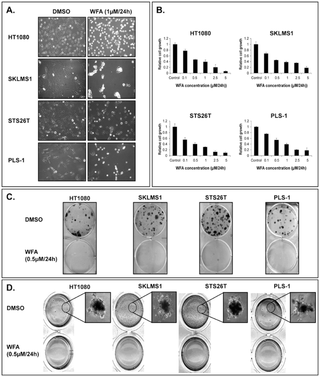
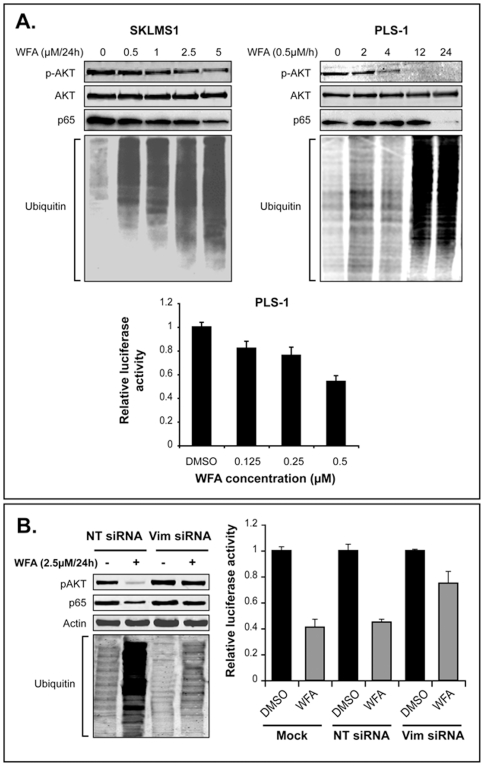
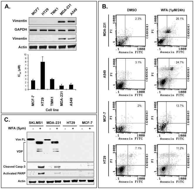
Background: Vimentin is a ubiquitous mesenchymal intermediate filament supporting mechano-structural integrity of quiescent cells while participating in adhesion, migration, survival, and cell signaling processes via dynamic assembly/disassembly in activated cells. Soft tissue sarcomas and some epithelial cancers exhibiting "epithelial to mesenchymal transition" phenotypes express vimentin. Withaferin-A, a naturally derived bioactive compound, may molecularly target vimentin, so we sought to evaluate its effects on tumor growth in vitro and in vivo thereby elucidating the role of vimentin in drug-induced responses.

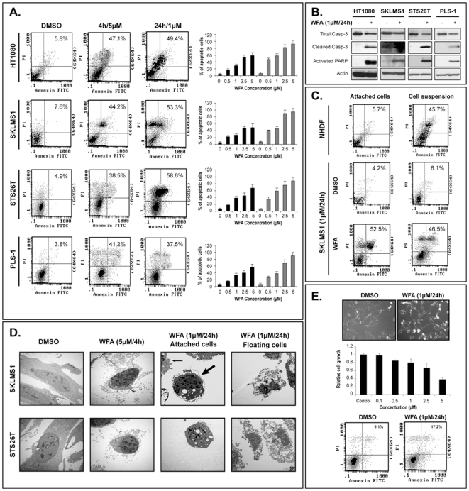
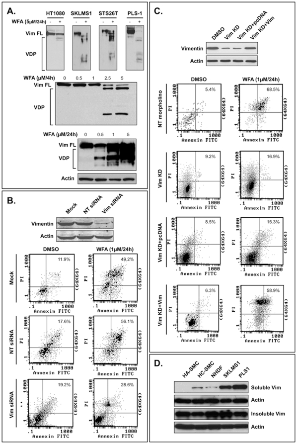
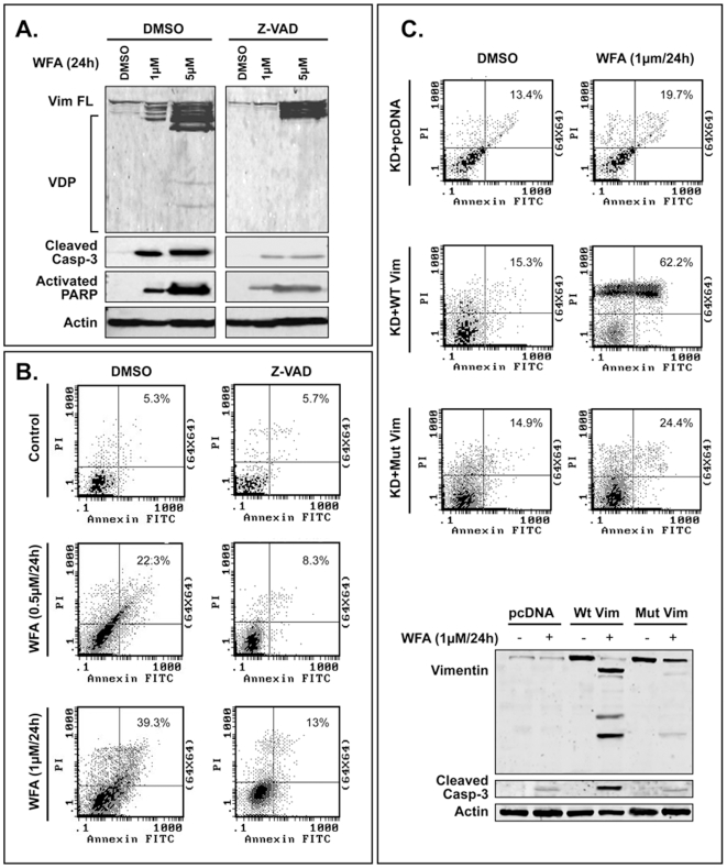
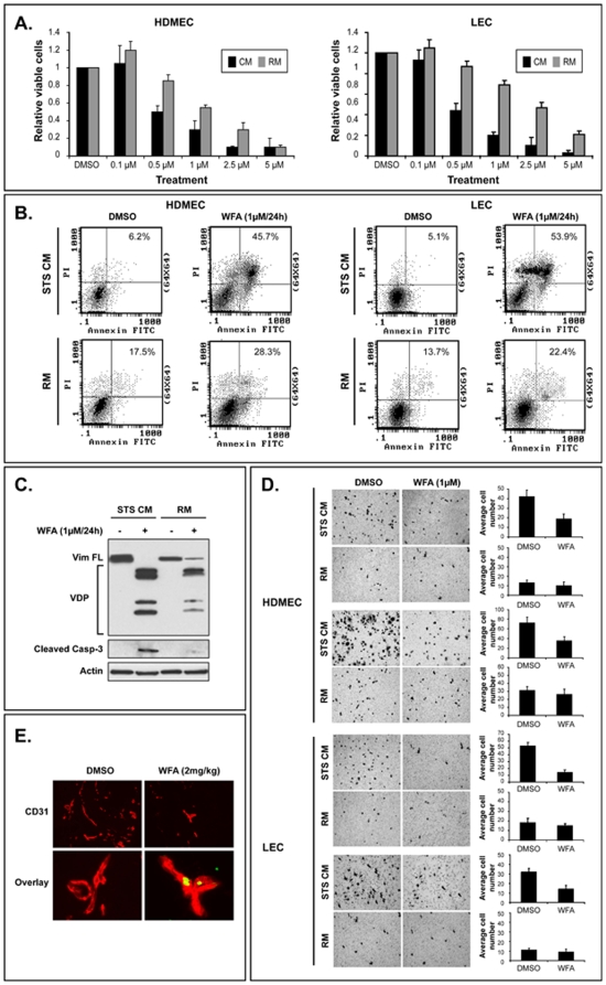
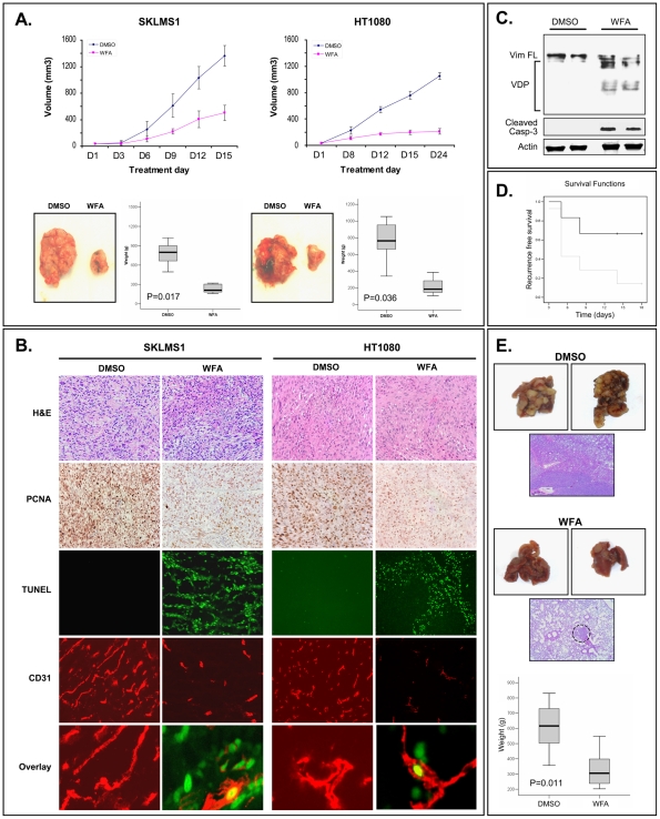
Methods and findings: Withaferin-A elicited marked apoptosis and vimentin cleavage in vimentin-expressing tumor cells but significantly less in normal mesenchymal cells. This proapoptotic response was abrogated after vimentin knockdown or by blockade of caspase-induced vimentin degradation via caspase inhibitors or overexpression of mutated caspase-resistant vimentin. Pronounced anti-angiogenic effects of Withaferin-A were demonstrated, with only minimal effects seen in non-proliferating endothelial cells. Moreover, Withaferin-A significantly blocked soft tissue sarcoma growth, local recurrence, and metastasis in a panel of soft tissue sarcoma xenograft experiments. Apoptosis, decreased angiogenesis, and vimentin degradation were all seen in Withaferin-A treated specimens.

Conclusions: In light of these findings, evaluation of Withaferin-A, its analogs, or other anti-vimentin therapeutic approaches in soft tissue sarcoma and "epithelial to mesenchymal transition" clinical contexts is warranted.


Conflict of interest statement

Competing Interests: The authors have declared that no competing interests exist.

Figures

Figure 1
Figure 1. WFA inhibits STS cell growth.
A) WFA treatment (1 µM/24 h) results in marked morphological changes, including cell-rounding and nuclear condensation, in STS cells; B) MTS assays demonstrate a WFA-induced, dose-dependent decrease in STS cell growth; C) WFA (0.5 µM/24 h) markedly inhibits STS cell colony formation capacity measured after ten days; D) WFA (0.5 µM/24 h) abrogates STS cell anchorage independent growth measured after three weeks. Graphs represent the average of three repeated experiments ±SD.
Figure 2
Figure 2. WFA induces marked apoptosis in STS cells.
A) Annexin-V/FACS analyses demonstrating marked WFA-induced apoptosis in STS cells (black bars represent 4 h of WFA treatment and gray bars represent 24 h); B) WFA (1 µM/24 hr) induces caspase-3 (Casp-3) cleavage and PARP activation in STS cells (WB analysis); C) STS cells are resistant to anoikis as compared to normal human dermal fibroblasts (NHDF). WFA (1 µM/24 hr) induces apoptosis in both attached and floating STS cells; D) Transmission electron microscopy (TEM) photographs depicting STS cell apoptosis (large arrow–nuclear condensation, small arrow–cytoplasmic blebbing) in response to WFA. Necrosis is demonstrated in floating STS cells; E) NHDF are more resistant to the effects of WFA (IC50: 3.7 µM±0.15) as compared to STS cells. Graphs represent the average of three repeated experiments ±SD.
Figure 3
Figure 3. WFA induces vimentin degradation and vimentin knockdown decreases cells' sensitivity to WFA.
A) WFA treatment (5 µM/4 h) results in decreased full-length (FL) vimentin levels and increased expression of vimentin degradation products (VDP) in all STS cells tested. A WFA dose dependent effect in SKLMS1 cells treated for 4 h is also shown. Vimentin cleavage is noticed secondary to low WFA concentrations after 24 h of treatment; B) Anti-vimentin SMARTpool siRNA (20 nM) elicits a marked decrease in vimentin expression in SKLMS1 cells (WB). Vimentin knockdown substantially blocks WFA-induced (1 µM/24 hr) apoptosis; C) Endogenous vimentin was first knocked down in SKLMS1 cells using anti-vimentin antisense phosphorodiamidate morpholino oligomers. After knockdown, vimentin was forcefully re-expressed in the cells (WB). Similar to the results of siRNA knockdown, anti-vimentin morpholino oligomers significantly blocks WFA-induced (1 µM/24 hr) apoptosis. Re-expression of vimentin restores SKLMS1 sensitivity to WFA; D) STS cells (SKLMS1 and PLS1) express significantly higher levels of soluble vimentin as compared to normal mesenchymal cells (smooth muscle cells: HA-SMC and HC-SMC and fibroblasts: NHDF). (NT siRNA  =  non targeting siRNA, Vim siRNA  =  anti-vimentin siRNA smartpool; NT morpholino  =  non targeting morpholino; Vim KD  =  vimentin knockdown)
Figure 4
Figure 4. WFA-induced vimentin degradation is caspase-dependent.
A) Pretreatement (4 h) of SKLMS1 cells with Z-VAD (a pan-caspase inhibitor) results in decreased WFA-induced (24 h) vimentin degradation in conjunction with lower levels of both cleaved caspase-3 and activated PARP; B) Z-VAD (4 h) pretreatment significantly abrogates WFA-induced (24 h) apoptosis in SKLMS1 cells; C) Vimentin knocked down SKLMS1 cells were transfected to express either wild-type vimentin or vimentin mutated at caspase cleavage sites (D85N and D259N). WFA (1 µM/24 hr) induces marked apoptosis in wild-type vimentin transfected cells in which vimentin degradation and caspase-3 activation can be detected (WB). However, a significant decrease in WFA-induced apoptosis in cells expressing the mutated vimentin, is noticed as well as a decrease in both vimentin degradation and caspase-3 activation. (WT Vim =  wild type vimentin; Mut Vim  =  mutated vimentin; Vim FL  =  full length vimentin; VDP  =  vimentin degradation products)
Figure 5
Figure 5. WFA-induced molecular deregulations are, at least partly, mediated by vimentin.
A) WFA treatment induces a dose- and time- dependent decrease in pAKT without effect on total AKT. Similarly, a dose-dependent decrease in NF-κB (p65) protein expression is also seen, although this decrease occurs only after 24 h of treatment. NF-κB activity in PLS1 cells, as depicted in graphs representing luciferase reporter assay results, is shown to be inhibited early after treatment (4 h). A marked dose- and time-dependent accumulation of ubiquitinated proteins is also shown; B) Vimentin knockdown in SKLMS1 cells abrogates WFA (2.5 µM/24 h)-induced pAKT inhibition, NF-κB protein decrease, and increased levels of protein ubiquitination. Similarly, vimentin knockdown blocks WFA (1 µM/4 h)-induced decrease in NF-κB activity. Graphs represent the average of three repeated experiments ±SD.
Figure 6
Figure 6. WFA induces apoptosis in endothelial cells cultured in STS-conditioned media.
A) A significantly higher rate of WFA-induced (24 h) growth inhibition is seen in endothelial cells (human dermal microvessel endothelial cells–HDMEC and murine lung endothelial cells–LEC) cultured in STS conditioned medium (CM) than in control regular medium (RM); B) WFA(1 µM/24 hr) induces higher levels of apoptosis in endothelial cells grown in STS-CM than in control medium; C) WFA (1 µM/24 hr) induces significantly higher rates of vimentin degradation and caspase-3 activation in endothelial cells grown in STS-CM than in quiescent endothelial cells; D) WFA (1 µM) abrogates migration and invasion of endothelial cells cultured in STS-CM; E) WFA (2 mg/kg) results in a significant decrease in the mean number of CD31-positive (red) blood vessels compared to control DMSO treatment in an in vivo gelfoam assay. CD-31(red)/TUNEL(green) double staining reveals endothelial cell apoptosis in WFA-treated mice. Graphs represent the average of three repeated experiments ±SD. (Vim FL  =  full length vimentin; VDP  =  vimentin degradation products)
Figure 7
Figure 7. Epithelial origin cancers' sensitivity to WFA is enhanced in cells exhibiting epithelial to mesenchymal transition (EMT).
A) WFA-induced growth inhibition corresponds to vimentin expression level in epithelial origin cancer cells; B) WFA (1 µM for 24 h) elicits a significantly higher apoptotic rate in vimentin-expressing carcinoma cells; C) WB analysis demonstrating vimentin degradation MDA231 cells in conjunction with enhanced cleaved caspase-3 and activated PARP expression levels. In contrast, only minimal or no expression of cleaved caspase-3 and activated PARP are seen in the vimentin-negative MCF7 and HT29 cells. Graphs represent the average of three repeated experiments ±SD. (Vim FL  =  full length vimentin; VDP  =  vimentin degradation products)
Figure 8
Figure 8. WFA abrogates STS growth, angiogenesis, recurrence, and metastasis in vivo.
A) WFA (2 mg/kg) significantly inhibits STS local growth and tumor weight; B) WFA treatment decreases tumor cell proliferation (PCNA), enhances apoptosis (TUNEL), and inhibits STS associated angiogenesis (CD31). CD31 (red)/TUNEL (green) double immunofluorescence analysis demonstrating apoptotic endothelial cells in WFA-treated tumors; C) WB analysis of tissue samples demonstrating vimentin degradation and caspase-3 activation in WFA-treated tumors; D) Kaplan Meier curves demonstrating a statistically significant (P = 0.02) delay in SKLMS1 local recurrence in WFA treated mice (black curve) compared to control (gray curve); E) Control mice (DMSO treated) exhibit numerous large MPNST lung metastases almost completely replacing the lung parenchyma. In contrast, WFA-treated mice exhibit markedly fewer microscopic small lung nodules (circle). WFA treatment significantly decreases average lung weights as compared to controls (box plots). (Vim FL  =  full length vimentin; VDP  =  vimentin degradation products).

References

    1. Yang J, Du X, Chen K, Ylipaa A, Lazar AJ, et al. Genetic aberrations in soft tissue leiomyosarcoma. Cancer Lett. 2009;275:1–8. - PMC - PubMed
    1. Fletcher CDM UK, editor. Pathology and Genetics of Soft Tissue and Bone. Lyon, France: IARC Press; 2002. World Health Organization Classification of Tumors.
    1. Sorensen PH, Lynch JC, Qualman SJ, Tirabosco R, Lim JF, et al. PAX3-FKHR and PAX7-FKHR gene fusions are prognostic indicators in alveolar rhabdomyosarcoma: a report from the children's oncology group. J Clin Oncol. 2002;20:2672–2679. - PubMed
    1. de Alava E, Kawai A, Healey JH, Fligman I, Meyers PA, et al. EWS-FLI1 fusion transcript structure is an independent determinant of prognosis in Ewing's sarcoma. J Clin Oncol. 1998;16:1248–1255. - PubMed
    1. Hirota S, Isozaki K, Moriyama Y, Hashimoto K, Nishida T, et al. Gain-of-function mutations of c-kit in human gastrointestinal stromal tumors. Science. 1998;279:577–580. - PubMed

Publication types