

A cis-regulatory signature for chordate anterior neuroectodermal genes
- PMID: 20419150
- PMCID: PMC2855326
- DOI: 10.1371/journal.pgen.1000912
A cis-regulatory signature for chordate anterior neuroectodermal genes
Abstract
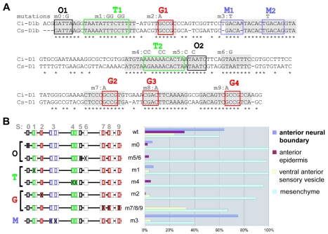
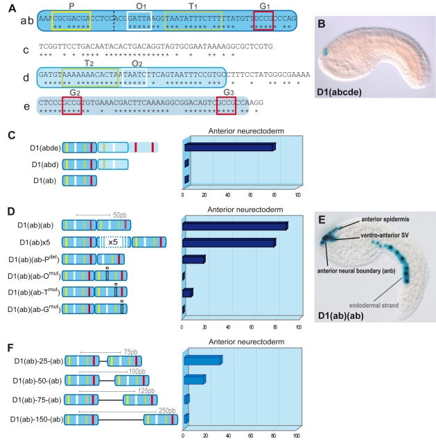
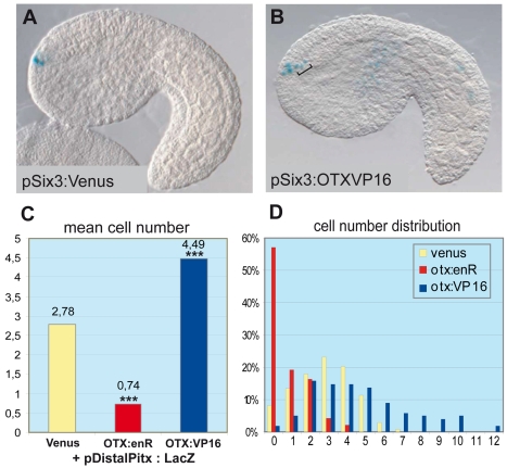
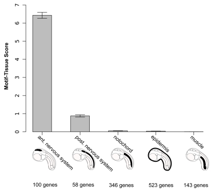
One of the striking findings of comparative developmental genetics was that expression patterns of core transcription factors are extraordinarily conserved in bilaterians. However, it remains unclear whether cis-regulatory elements of their target genes also exhibit common signatures associated with conserved embryonic fields. To address this question, we focused on genes that are active in the anterior neuroectoderm and non-neural ectoderm of the ascidian Ciona intestinalis. Following the dissection of a prototypic anterior placodal enhancer, we searched all genomic conserved non-coding elements for duplicated motifs around genes showing anterior neuroectodermal expression. Strikingly, we identified an over-represented pentamer motif corresponding to the binding site of the homeodomain protein OTX, which plays a pivotal role in the anterior development of all bilaterian species. Using an in vivo reporter gene assay, we observed that 10 of 23 candidate cis-regulatory elements containing duplicated OTX motifs are active in the anterior neuroectoderm, thus showing that this cis-regulatory signature is predictive of neuroectodermal enhancers. These results show that a common cis-regulatory signature corresponding to K50-Paired homeodomain transcription factors is found in non-coding sequences flanking anterior neuroectodermal genes in chordate embryos. Thus, field-specific selector genes impose architectural constraints in the form of combinations of short tags on their target enhancers. This could account for the strong evolutionary conservation of the regulatory elements controlling field-specific selector genes responsible for body plan formation.
Conflict of interest statement
LC joined the faculty of New York University after the review process had commenced.
Figures
Similar articles
-
Making very similar embryos with divergent genomes: conservation of regulatory mechanisms of Otx between the ascidians Halocynthia roretzi and Ciona intestinalis.Development. 2005 Apr;132(7):1663-74. doi: 10.1242/dev.01707. Epub 2005 Mar 2. Development. 2005. PMID: 15743880
-
Evolution of anterior Hox regulatory elements among chordates.BMC Evol Biol. 2011 Nov 15;11:330. doi: 10.1186/1471-2148-11-330. BMC Evol Biol. 2011. PMID: 22085760 Free PMC article.
-
A cis-regulatory signature in ascidians and flies, independent of transcription factor binding sites.Curr Biol. 2010 May 11;20(9):792-802. doi: 10.1016/j.cub.2010.03.063. Epub 2010 Apr 29. Curr Biol. 2010. PMID: 20434338
-
Cis-regulation and conserved non-coding elements in amphioxus.Brief Funct Genomics. 2012 Mar;11(2):118-30. doi: 10.1093/bfgp/els006. Epub 2012 Mar 7. Brief Funct Genomics. 2012. PMID: 22402505 Review.
-
Gene-regulatory networks in the Ciona embryos.Brief Funct Genomic Proteomic. 2009 Jul;8(4):250-5. doi: 10.1093/bfgp/elp018. Epub 2009 Jun 17. Brief Funct Genomic Proteomic. 2009. PMID: 19535506 Review.
Cited by
-
Initial deployment of the cardiogenic gene regulatory network in the basal chordate, Ciona intestinalis.Dev Biol. 2012 Aug 1;368(1):127-39. doi: 10.1016/j.ydbio.2012.05.002. Epub 2012 May 14. Dev Biol. 2012. PMID: 22595514 Free PMC article.
-
A transiently expressed connexin is essential for anterior neural plate development in Ciona intestinalis.Development. 2013 Jan 1;140(1):147-55. doi: 10.1242/dev.084681. Epub 2012 Nov 22. Development. 2013. PMID: 23175630 Free PMC article.
-
Study of Cis-regulatory Elements in the Ascidian Ciona intestinalis.Curr Genomics. 2013 Mar;14(1):56-67. doi: 10.2174/138920213804999192. Curr Genomics. 2013. PMID: 23997651 Free PMC article.
-
Brachyury, Foxa2 and the cis-Regulatory Origins of the Notochord.PLoS Genet. 2015 Dec 18;11(12):e1005730. doi: 10.1371/journal.pgen.1005730. eCollection 2015 Dec. PLoS Genet. 2015. PMID: 26684323 Free PMC article.
-
ACAM, a novel member of the neural IgCAM family, mediates anterior neural tube closure in a primitive chordate.Dev Biol. 2016 Jan 1;409(1):288-296. doi: 10.1016/j.ydbio.2015.10.032. Epub 2015 Nov 2. Dev Biol. 2016. PMID: 26542009 Free PMC article.
References
-
- Garcia-Bellido A. Genetic control of wing disc development in Drosophila. Ciba Found Symp. 1975;0:161–182. - PubMed
-
- Blackshaw S, Fraioli RE, Furukawa T, Cepko CL. Comprehensive analysis of photoreceptor gene expression and the identification of candidate retinal disease genes. Cell. 2001;107:579–589. - PubMed
-
- Chen S, Wang QL, Nie Z, Sun H, Lennon G, et al. Crx, a novel Otx-like paired-homeodomain protein, binds to and transactivates photoreceptor cell-specific genes. Neuron. 1997;19:1017–1030. - PubMed
-
- Hsiau TH, Diaconu C, Myers CA, Lee J, Cepko CL, et al. The cis-regulatory logic of the mammalian photoreceptor transcriptional network. PLoS ONE. 2007;2:e643. doi: 10.1371/journal.pone.0000643. - DOI - PMC - PubMed
Publication types
MeSH terms
Substances
LinkOut - more resources
Full Text Sources
