. 2010:619:339-63.
doi: 10.1007/978-1-60327-412-8_20.

In vitro dissection of protein translocation into the mammalian endoplasmic reticulum

Affiliations

In vitro dissection of protein translocation into the mammalian endoplasmic reticulum

Ajay Sharma et al. Methods Mol Biol. 2010.

Abstract

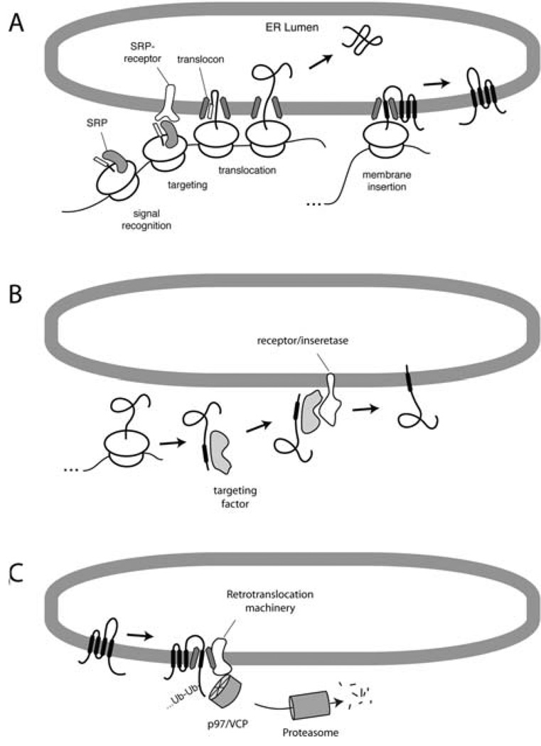
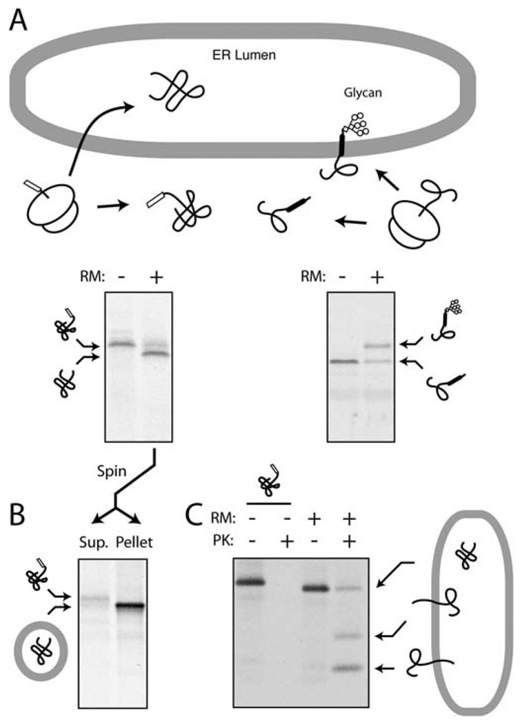
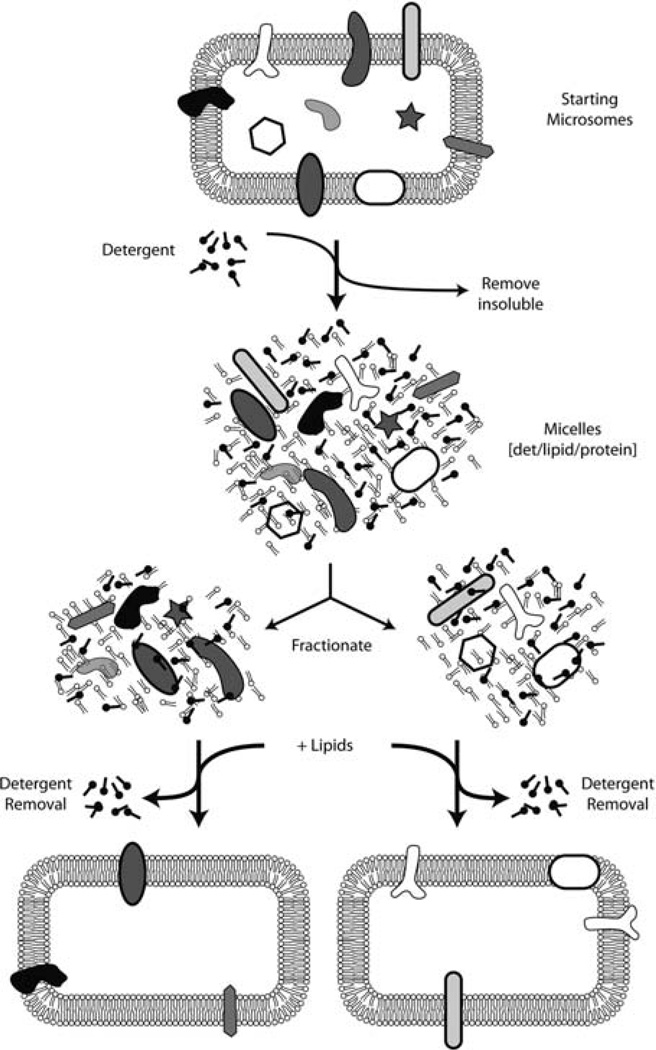
In eukaryotic cells, roughly one-fourth of all mRNAs code for secretory and membrane proteins. This class of proteins must first be segregated to the endoplasmic reticulum, where they are either translocated into the lumen or inserted into the lipid bilayer. The study of these processes has long relied on their successful reconstitution in cell-free systems. The high manipulability of such in vitro systems has allowed the identification of key machinery, elucidation of their functional roles in translocation, and dissection of their mechanisms of action. Here, we provide the basic methodology for (i) setting up robust mammalian-based in vitro translation and translocation systems, (ii) assays for protein translocation, insertion, and topology, and (iii) methods to solubilize, fractionate, and reconstitute ER membranes. Variations of these methods should be applicable not only to forward protein translocation systems but also for dissecting other poorly understood membrane-associated processes such as retrotranslocation.


Figures

Fig. 20.1
Fig. 20.1
Examples of ER-associated pathways amenable to in vitro reconstitution. (A) The SRP-dependent co-translational translocation pathway. (B) A post-translational translocation pathway for tail-anchored membrane protein insertion. (C) Post-translocational pathway of membrane protein metabolism involving ubiquitination, retrotranslocation, and proteasomal degradation. In each of these instances, the substrate is synthesized in vitro, making it the only protein that becomes radiolabeled. The other components of the system can be manipulated to analyze the requirements for substrate translocation, maturation, degradation, etc.
Fig. 20.2
Fig. 20.2
Assays for protein segregation to the ER. (A) ER translocation-dependent modification. In the example on the left, a precursor becomes processed by signal peptidase only upon its translocation into the lumen of rough microsomes (RM), an event that can be monitored by a change in migration on SDS-PAGE. In the example on the right, a glycosylation site becomes modified upon successful translocation (8). (B) Co-fractionation assays. A sample similar to that from panel A can be separated by centrifugation into a cytosolic supernatant and membrane pellet to assess successful translocation. (C) Protease protection assay. Upon addition of proteinase K (PK) to the products of a translocation reaction, proteins that are either fully or partially translocated into the lumen of RMs are protected. Even a protein that generates multiple topological forms (such as mammalian prion protein; see ref. 20) can be resolved by this assay. By contrast, lack of translocation leads to complete digestion upon PK treatment.
Fig. 20.3
Fig. 20.3
Schematic depiction of membrane protein reconstitution. Crude microsomes are solubilized with detergent, fractionated, and reconstituted into proteoliposomes by removal of detergent in the presence of phospholipids. Note that not all proteins are successfully reconstituted, and the orientation achieved after reconstitution must be checked empirically.
Fig. 20.4
Fig. 20.4
Example of differential solubilization and reconstitution of membrane proteins. Crude RMs (lane 1) were sequentially extracted by four buffers containing different amounts of detergent and salt to generate four supernatant fractions (S1–S4) and an insoluble pellet (primarily containing ribosomes). Each of these four fractions was then reconstituted in the presence of phospholipids by detergent removal, and the resulting proteoliposomes were also analyzed on the gel. Note that the very abundant high molecular weight proteins in S1 and S2 (primarily lumenal proteins) are not reconstituted. Note also the different protein profiles of the different proteoliposomes illustrating the utility of differential membrane protein extraction as a purification step. The detergent in this case was DeoxyBigCHAP, although similar results can be obtained with other detergents.

References

    1. Walter P, Blobel G. Preparation of microsomal membranes for cotranslational protein translocation. Methods Enzymol. 1983;96:84–93. - PubMed
    1. Adelman MR, Blobel G, Sabatini DD. An improved cell fractionation procedure for the preparation of rat liver membrane-bound ribosomes. J Cell Biol. 1973;56:191–205. - PMC - PubMed
    1. Wilson R, Allen AJ, Oliver J, Brookman JL, High S, Bulleid NJ. The translocation, folding, assembly and redox-dependent degradation of secretory and membrane proteins in semi-permeabilized mammalian cells. Biochem J. 1995;307:679–687. - PMC - PubMed
    1. Stefanovic S, Hegde RS. Identification of a targeting factor for posttranslational membrane protein insertion into the ER. Cell. 2007;128:1147–1159. - PubMed
    1. Schuldiner M, Metz J, Schmid V, Denic V, Rakwalska M, Schmitt HD, Schwappach B, Weissman JS. The GET complex mediates insertion of tail-anchored proteins into the ER membrane. Cell. 2008;134:634–645. - PMC - PubMed

Publication types

Substances

LinkOut - more resources