

doi: 10.1007/978-1-60327-412-8_24.
High-quality immunofluorescence of cultured cells
Affiliations
- PMID: 20419424
- PMCID: PMC2893412
- DOI: 10.1007/978-1-60327-412-8_24
Item in Clipboard
High-quality immunofluorescence of cultured cells
Methods Mol Biol.
2010.
Abstract
Immunofluorescence microscopy of cultured cells often gives poor preservation of delicate structures. We have obtained dramatically improved results with a simple modification of a standard protocol. Cells growing on a coverslip are rapidly dehydrated in a cold organic solvent and then are rehydrated in a solution containing a homobifunctional crosslinker. The crosslinking reaction stabilizes cellular structures during subsequent incubation and wash steps, usually without compromising antigenicity. This method reproducibly yields high-quality images of endomembrane compartments and cytoskeletal elements.
Figures
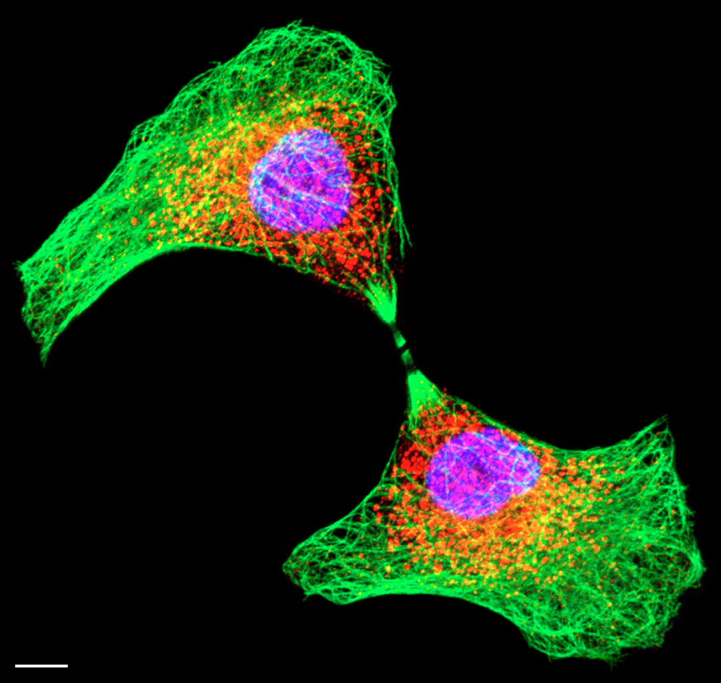
Immunofluorescence image of a dividing normal rat kidney cell. Microtubules (green) were stained with a monoclonal anti-β-tubulin antibody (clone KMX-1, Roche, Indianapolis, IN) followed by Cy2-conjugated donkey antimouse antibody. tER sites (red) were stained with an affinity-purified polyclonal anti-Sec13 antibody [6] followed by Rhodamine Red-X-conjugated donkey anti-rabbit antibody. Both primary antibodies were diluted 1:100, and both secondary antibodies (from Jackson Immunoresearch, West Grove, PA) were diluted 1:200. DNA (magenta) was stained by supplementing the mounting medium with 4 mM TOTO-3 (Molecular Probes, Eugene, OR). Separate Z-stacks in three fluorescence channels were collected with a Zeiss (Thornwood, NY) LSM 510 confocal microscope equipped with a 100X 1.4-NA Plan-Apo objective and with standard filters for visualizing FITC/Cy2, Rhodamine Red-X, and Cy5/TOTO-3. These images were then projected and combined using the Zeiss software. The background staining outside of the cells was removed using Adobe Photoshop. Scale bar, 10 µm.
References
-
- Donaldson JG. Current Protocols in Cell Biology. John Wiley & Sons; 1998. Immunofluorescence Staining; pp. 4.3.1–4.3.6. - PubMed
-
- Melan MA, Sluder G. Redistribution and differential extraction of soluble proteins in permeabilized cultured cells. Implications for immunofluorescence microscopy. J. Cell Sci. 1992;101:731–743. - PubMed
-
- Staudt T, Lang MC, Medda R, Engelhardt J, Hell SW. 2,2'-Thiodiethanol: a new water soluble mounting medium for high resolution optical microscopy. Microsc. Res. Tech. 2007;70:1–9. - PubMed
Publication types
MeSH terms
Grants and funding
LinkOut - more resources
Full Text Sources
