. 2010 May;239(5):1555-72.
doi: 10.1002/dvdy.22274.

PRP-17 and the pre-mRNA splicing pathway are preferentially required for the proliferation versus meiotic development decision and germline sex determination in Caenorhabditis elegans

Affiliations

PRP-17 and the pre-mRNA splicing pathway are preferentially required for the proliferation versus meiotic development decision and germline sex determination in Caenorhabditis elegans

Jessica Amrozowicz Kerins et al. Dev Dyn. 2010 May.

Abstract

In C. elegans, the decision between germline stem cell proliferation and entry into meiosis is controlled by GLP-1 Notch signaling, which promotes proliferation through repression of the redundant GLD-1 and GLD-2 pathways that direct meiotic entry. We identify prp-17 as another gene functioning downstream of GLP-1 signaling that promotes meiotic entry, largely by acting on the GLD-1 pathway, and that also functions in female germline sex determination. PRP-17 is orthologous to the yeast and human pre-mRNA splicing factor PRP17/CDC40 and can rescue the temperature-sensitive lethality of yeast PRP17. This link to splicing led to an RNAi screen of predicted C. elegans splicing factors in sensitized genetic backgrounds. We found that many genes throughout the splicing cascade function in the proliferation/meiotic entry decision and germline sex determination indicating that splicing per se, rather than a novel function of a subset of splicing factors, is necessary for these processes.


Figures

Figure 1
Figure 1. The prp-17(oz273) phenotype
Fluorescent images of single adult hermaphrodite gonad arms that have been dissected away from the body. The top image in each panel depicts DAPI staining (blue) to visualize DNA morphology. The bottom image shows the proliferative cell nuclei (green) detected with antibodies specific to REC-8, while meiotic prophase nuclei (red) are detected with HIM-3 (see Hansen et al. 2004b). Animals were grown continuously at 20°C. Distal end is to the left. Scale bar = 20 μm. (A) Wild-type hermaphrodite. The ~20 cell diameter proliferative zone consists of REC-8-positive, HIM-3-negative germ cells, whereas cells that have entered meiotic prophase are HIM-3-positive, REC-8-negative. Proliferation, meiotic prophase, the loop region (where gametogenesis begins, dotted line), and oocytes are labeled. (B) The glp-1(oz264) hermaphrodite at 20° appears wild-type. (C) prp-17(oz273) mutants are Mog, characterized by an absence of oocytes and a vast excess sperm in the proximal gonad and 1° spermatocytes (Spc) in the loop region and extending into the distal gonad. (D) prp-17(oz273);glp-1(oz264) hermaphrodite has a tumorous germline, with REC-8-positive cells throughout most of the gonad.
Figure 2
Figure 2. prp-17 and HTATSF1 function in meiotic entry and sex determination
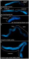
Fluorescent images of single adult hermaphrodite gonad arms that have been dissected away from the body and stained with DAPI to visualize DNA morphology. Distal end is to the left. Scale bar = 20 μm. (A) The fog-3(q443) prp-17(oz273) hermaphrodite does not make sperm, but does make oocytes (shown in proximal region), although abnormal. (B) prp-17(oz273); gld-3(q730) hermaphrodites show a late-onset tumor and proximal proliferation, indicated by DAPI staining (regions of proliferation are noted, based on REC-8/HIM-3 staining, which is not shown). (Actual genotype: prp-17(oz273); gld-3(q730); unc-32(e189)) (C) prp-17(oz273); gld-3(q730); glp-1(q175) hermaphrodites are tumorous, showing proliferating cells throughout the germline. (Actual genotype: prp-17(oz273); gld-3(q730); unc-32(e189) glp-1(q175)) (D) and (E) Y65B4A.1(RNAi) (human ortholog: HTATSF1) phenocopies prp-17(oz273) in both glp-1(+) and glp-1(oz264) genetic backgrounds. (D) rrf-1(pk1417); Y65B4A.1(RNAi) hermaphrodites are Mog, exhibiting excess sperm, 1° spermatocytes (Spc) and no oocytes. (E) rrf-1(pk1417); glp-1(oz264); Y65B4A.1(RNAi) animals show overproliferation throughout the germline.
Figure 3
Figure 3. The prp-17 gene and gene product
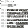
(A) The prp-17 message contains 4 exons (boxes). 5′ and 3′ UTRs are shown in black. The oz273 mutation deletes the first 521 base pairs of exon 2. (B) Top: the predicted PRP-17 protein contains 567 amino acids. The main functional motifs, six WD-40 repeats, are shown in black. The hatched box indicates a 4-amino acid motif shown to be essential for splicing in yeast (Lindsey-Boltz et al, 2000) (see C). Bottom: the predicted truncated protein produced in prp-17(oz273) mutants. The oz273 deletion mutation creates a premature STOP codon upstream of all functional motifs. The shaded box indicates aberrant amino acids (and premature STOP codon) created by the predicted cryptic splice site of the mutant. (C) Alignment of the conserved C-terminal 221–567 amino acids of the C. elegans PRP-17 protein (accession no. NP_492851.1) with yeast (S. cerevisiae, accession no. NP_010652.1), human (accession no. NP_056975.1), and fly (D. melanogaster, accession no. NP_651005.1) orthologs. Residues in black are identical in two or more orthologs. WD-40 repeats are underlined. Asterisks denote the 4-amino acid motif described in (B).
Figure 4
Figure 4. Complementation of a yeast PRP17 knockout strain
The yeast strain prp17Δ BJ2168 was transformed with the high copy plasmid pG1 alone or with pG1 containing full-length yPRP17, hPRP17, or CePRP17. 10-fold serial dilutions were made of each transformation, which were then grown on medium lacking tryptophan for 3–6 days at 25, 30, and 37°C. The figure shows 1:100 dilutions of each transformation grown for 4 days at each temperature.
Figure 5
Figure 5. The pre-mRNA splicing pathway and C. elegans genes that function in the proliferation/meiotic development decision and germline sex determination
Adapted from Ohi et al. 2005. Schematic drawing of the pre-mRNA splicing pathway. The assembly of an activated spliceosome onto a nascent transcript is necessary for pre-mRNA splicing to occur. Assembly begins when U1 and U2 bind to the 5′ and 3′ splice sites, respectively (Complexes E and A). The U5/U4/U6 tri-snRNP then binds (Complex B), followed by rearrangement of the spliceosomal complex which releases U1 and U4 (Complex B*). The first catalytic step of splicing occurs next, resulting in the release of the intron at the 5′ splice site, and subsequent formation of the lariat intermediate (Complex C). The second catalytic step then occurs, completely excising the intron and associated splicing complexes (post-splicing complex), resulting in fully spliced exons, which then undergo further processing and export from the nucleus. The post-splicing complex undergoes disassembly, and the cycle can begin again. The C. elegans splicing factor orthologs that display overproliferation and/or Mog phenotypes (Table 2) are listed near the step(s) where the yeast/human proteins have been shown to act (boxes), or listed in circles (to indicate those that function in processes not shown). YTD = the splicing step(s) in which these genes function is yet to be determined.
Figure 6
Figure 6. Genetic models for PRP-17 and splicing factors in the C. elegans proliferation/meiotic development decision and germline sex determination
Positive interactions are depicted with arrows, while negative interactions are depicted with T-bars. Proteins are shown in capital letters; gene names are shown in lower case italics. (A) The core GLP-1 Notch signaling pathway shown with the downstream GLD-1 and GLD-2 pathways that promote entry into meiosis (Kimble and Crittenden 2005; Hansen and Schedl 2006). PRP-17 and other C. elegans splicing factor orthologs promote meiotic development by positively regulating the GLD-1 pathway through the splicing of mRNAs of genes that function in this pathway. Because the relevant direct splicing targets of PRP-17/ spliceosome have not yet been identified, we show general positive regulation on the GLD-1 pathway as a whole. (B) The core germline sex determination pathway in C. elegans (Ellis and Schedl 2006). In hermaphrodites GLD-1 and FOG-2 act to promote spermatogenesis by repression of tra-2 mRNA in early larvae; the switch to oogenesis occurs upon repression of fem-3 mRNA and derepression of tra-2 mRNA in late larvae. PRP-17 and other splicing factor orthologs may promote the oocyte fate in C. elegans germline sex determination via splicing of the mRNA of an unknown gene (or genes) X, which functions as a translational repressor of fem-3 mRNA during hermaphrodite oogenesis. In (A), PRP-17 and the splicing factors have been placed downstream of GLP-1 signaling as triple mutants of glp-1 null, gld-3 null with prp-17, mog-1 or mog-6 mutations (described here) and teg-4 (Mantina et al. 2009) are tumorous, demonstrating that glp-1 activity is not required for the overproliferation phenotype. Previous work indicated that the GLD-2 polyA polymerase and the GLD-3 Bicaudal-C related RNA binding protein function together to promote entry into meiosis (Kadyk and Kimble,1998; Wang et al. 2002; Eckmann et al. 2004; Hansen et al. 2004a and b). Therefore, we were surprised to find that the gld-2(q497) prp-17(oz273); glp-1(q175) triple mutant is not tumorous, but rather has the glp-1 premature meiotic entry phenotype, with similar results in triple mutants with mog-1 and mog-6 (data not shown) as well as for teg-4 (Mantina et al. 2009). There are other situations where gld-2 and gld-3 do not behave identically in the proliferation versus meiosis decision. gld-3(q730) has a stronger meiotic entry defect in combination with gld-1 or nos-3 null alleles than does gld-2(q497) (Eckmann et al. 2004; Hansen et al. 2004a and b). The genetic behavior of the pas-5 proteasome subunit shows a similar set of interactions with gld-2, gld-3 and glp-1 as the splicing factor mutants: the pas-5; gld-3; glp-1 mutant is tumorous while gld-2 pas-5; glp-1 mutant shows the glp-1 premature meiotic entry phenotype (MacDonald et al. 2008). How might GLD-2 and GLD-3 function together yet display some different genetic behaviors? One possibility is that GLD-3 may have a separate function, outside of its role with GLD-2, in promoting entry into meiosis, possibly in the proposed third pathway that acts in parallel to the GLD-1 and GLD-2 pathways (Hansen et al. 2004b). Alternatively, although the gld-2(q497) allele used these experiments is a stop mutation (Wang et al. 2002), it may not fully eliminate gld-2 activity. In contrast, the gld-3(q730) allele is a deletion that likely eliminates all gld-3 activity. While the reasons for differences in behavior of gld-2 and gld-3 remain to be resolved, the finding that the tumorous phenotype of the gld-3 null with splicing factor mutants is independent of glp-1 activity strongly supports the proposal that the splicing factors function, at least in part, downstream of GLP-1 signaling.

Similar articles

Cited by

References

    1. Ahringer J, Kimble J. Control of the sperm-oocyte switch in Caenorhabditis elegans hermaphrodites by the fem-3 3′ untranslated region. Nature. 1991;349:346–348. - PubMed
    1. Ahringer J, Rosenquist TA, Lawson DN, Kimble J. The C. elegans gene fem-3 is regulated post-transcriptionally. EMBO J. 1992;11:2303–2310. - PMC - PubMed
    1. Austin J, Kimble J. glp-1 is required in the germ line for regulation of the decision between mitosis and meiosis in C. elegans. Cell. 1987;51:589–599. - PubMed
    1. Austin J, Kimble J. Transcript analysis of glp-1 and lin-12, homologous genes required for cell interaction during development of Caenorhabditis elegans. Cell. 1989;58:565–571. - PubMed
    1. Barton MK, Kimble J. fog-1, a regulatory gene required for specification of spermatogenesis in the germ line of Caenorhabditis elegans. Genetics. 1990;125:29–39. - PMC - PubMed

Publication types