. 2010 May 25;49(20):4308-17.
doi: 10.1021/bi1002585.

Mutation of Asn28 disrupts the dimerization and enzymatic activity of SARS 3CL(pro)

Affiliations

Mutation of Asn28 disrupts the dimerization and enzymatic activity of SARS 3CL(pro)

Jennifer Barrila et al. Biochemistry. .

Abstract

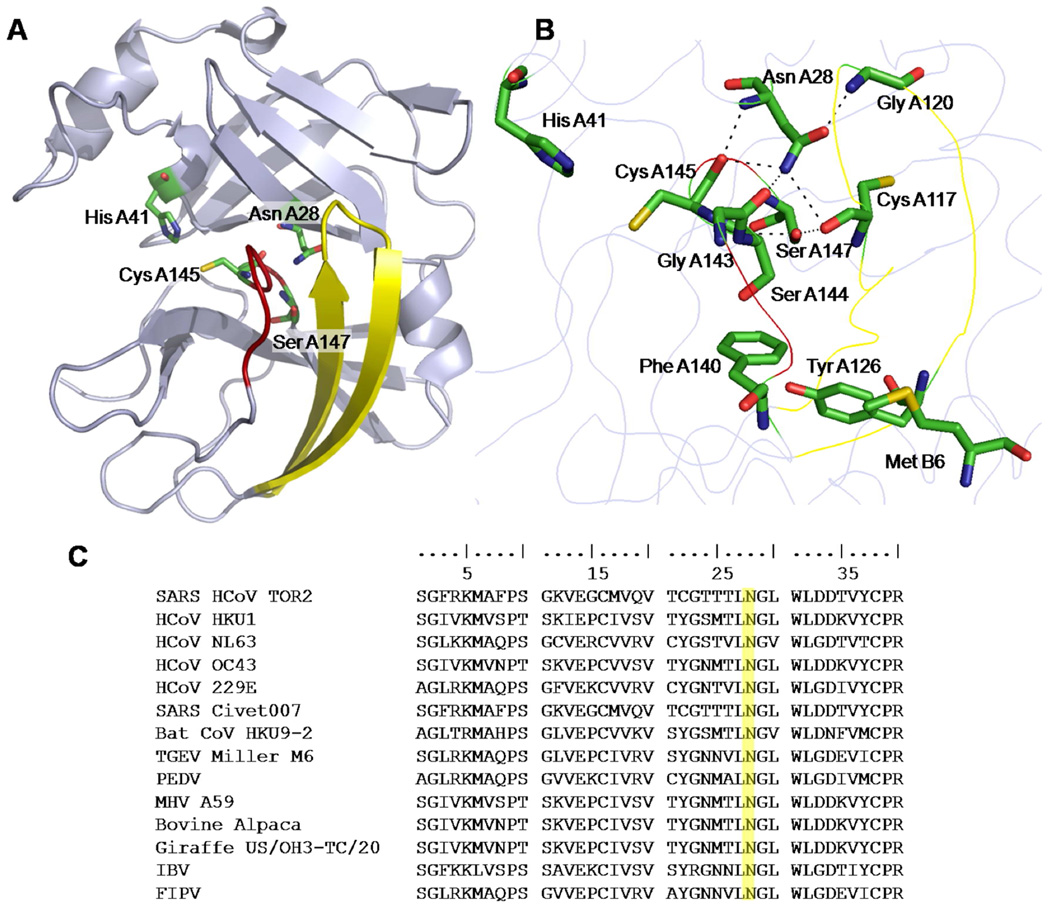
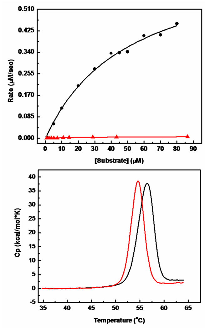
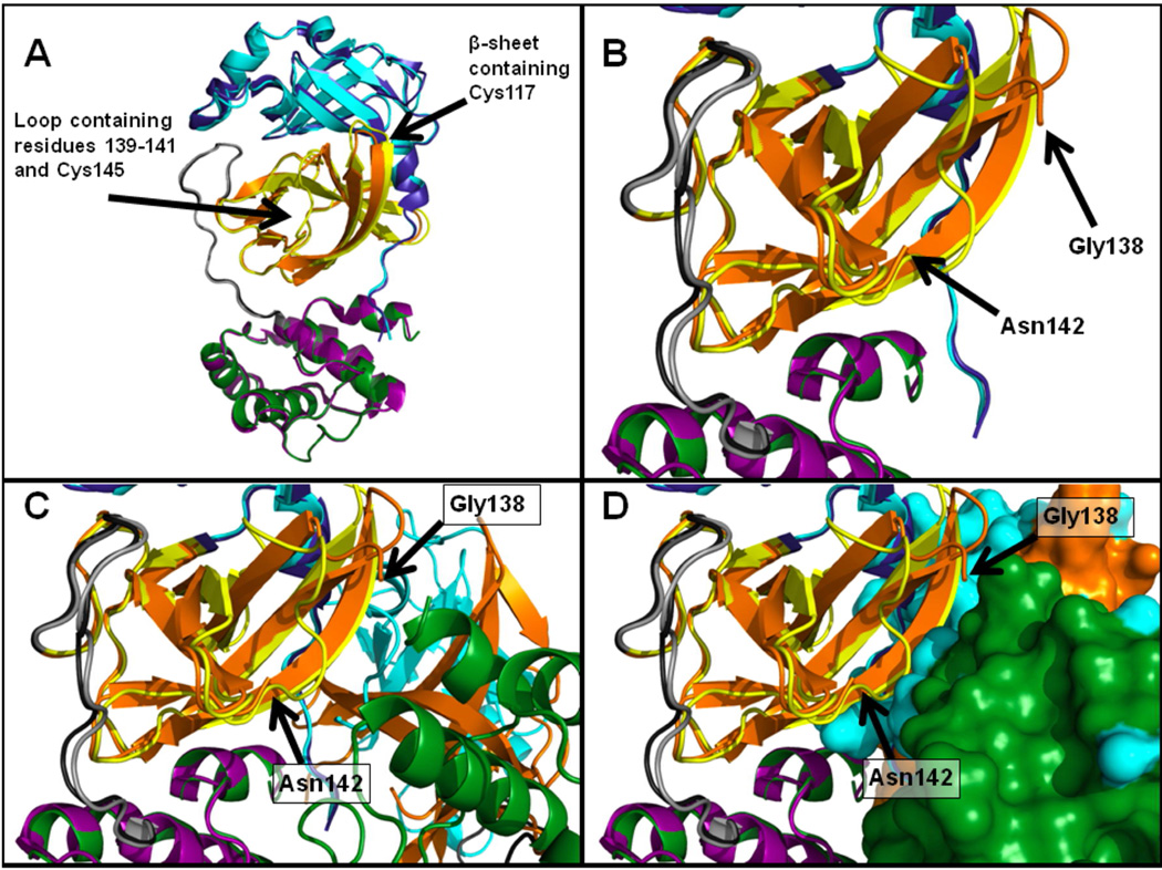
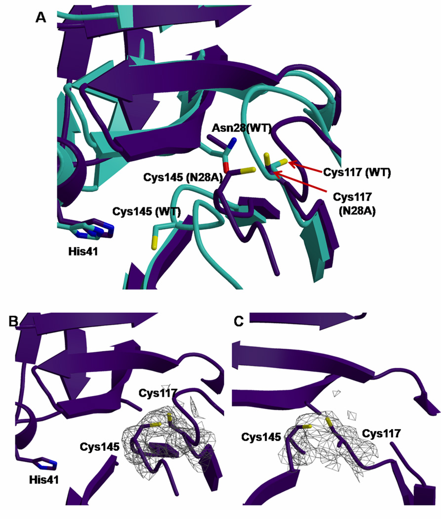
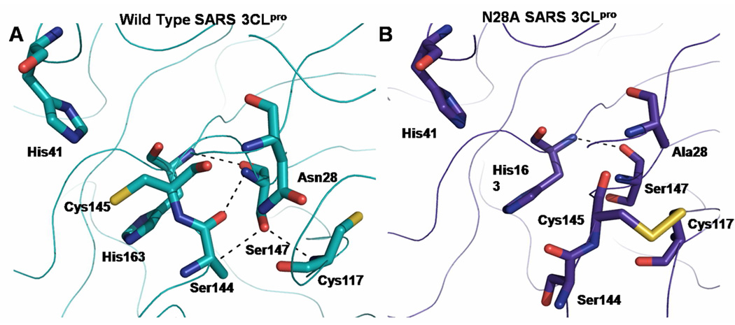
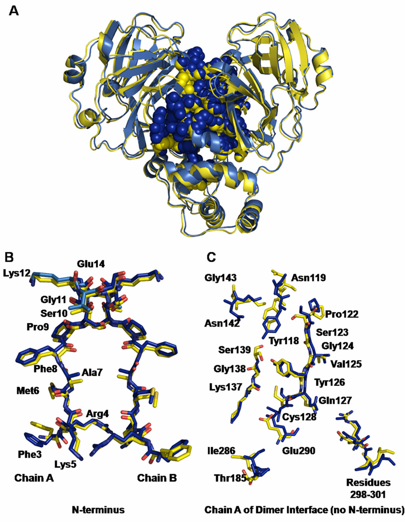
Coronaviruses are responsible for a significant proportion of annual respiratory and enteric infections in humans and other mammals. The most prominent of these viruses is the severe acute respiratory syndrome coronavirus (SARS-CoV) which causes acute respiratory and gastrointestinal infection in humans. The coronavirus main protease, 3CL(pro), is a key target for broad-spectrum antiviral development because of its critical role in viral maturation and high degree of structural conservation among coronaviruses. Dimerization is an indispensable requirement for the function of SARS 3CL(pro) and is regulated through mechanisms involving both direct and long-range interactions in the enzyme. While many of the binding interactions at the dimerization interface have been extensively studied, those that are important for long-range control are not well-understood. Characterization of these dimerization mechanisms is important for the structure-based design of new treatments targeting coronavirus-based infections. Here we report that Asn28, a residue 11 A from the closest residue in the opposing monomer, is essential for the enzymatic activity and dimerization of SARS 3CL(pro). Mutation of this residue to alanine almost completely inactivates the enzyme and results in a 19.2-fold decrease in the dimerization K(d). The crystallographic structure of the N28A mutant determined at 2.35 A resolution reveals the critical role of Asn28 in maintaining the structural integrity of the active site and in orienting key residues involved in binding at the dimer interface and substrate catalysis. These findings provide deeper insight into complex mechanisms regulating the activity and dimerization of SARS 3CL(pro).


Figures

Figure 1
Figure 1. Location of Asn28 within the catalytic domains of wild type SARS 3CLpro
(A) SARS 3CLpro catalytic domains (residues 8–184) [pdb file 2BX4 (18)] are depicted in ribbon representation. The loop containing residues 140–147 is colored red and the β-sheet containing residues 111–128 in yellow. Residues Asn28, His41, Cys145, and Ser147 are shown as sticks and colored by atom type with carbon in green, nitrogen in blue, oxygen in red and sulfur in yellow. (B) Key residues surrounding Asn28 are depicted as sticks and colored by atom type as in (A). Hydrogen bonding interactions made by Asn28 and Ser147 were calculated using Chimera (47) and are indicated by black dotted lines. (C) Asn28 is highly conserved among coronavirus 3CL proteases. The sequence alignment of fourteen coronavirus 3CL proteases was produced using ClustalW (48). The first forty residues are shown. Asn28 is highlighted in yellow in each of the sequences. Genbank accession numbers for protein sequences are as follows: SARS-CoV (TOR2 strain): AAP41036; HCoV HKU1 (genotype A): AY597011; HCoV NL63: AY567487; HCoV OC43: AY903460; HCoV 229E: AF304460, SARS Civet (palm civet isolate 007): AAU04645; Bat CoV (strain HKU9-2): AAZ41328; TGEV (Miller M6 strain): ABG89299, PEDV: NP_598309; MHV A59 (A59 strain): NP_045298; BCV Alpaca (Alpaca strain): ABI93997; Giraffe CoV (strain US/OH3-TC/2006); IBV: AAY24431; FIPV: ABI14447.
Figure 2
Figure 2. Enzymatic activities of wild type SARS 3CLpro versus the N28A mutant
(A) The enzymatic activities of wild type 3CLpro (●) and N28A (▲) were determined at 25°C with an enzyme concentration of 1 µM in 10mM sodium phosphate, 10mM NaCl, 1mM EDTA, 1mM TCEP (pH 7.4). The initial velocity (µM per second) is plotted as a function of substrate concentration. (B) Thermal denaturation of wild type (black) and N28A (red) SARS 3CLpro as determined by differential scanning calorimetry. Excess heat capacity is plotted as a function of temperature. Calorimetric scans for both proteins were performed at identical concentrations (0.2 mg/mL) at a scanning rate of 1°C per minute.
Figure 3
Figure 3. Sedimentation velocity and sedimentation equilibrium ultracentrifugation of N28A at 0.5 mg/ml
(A) Sedimentation velocity absorbance trace of N28A SARS 3CLpro at 280 nm. The sedimentation of the N28A mutant was carried out with a Beckman Coulter XL-I analytical ultracentrifuge at 20°C and 50,000 rpm. (B) Residuals of the experimental fit of N28A SARS 3CLpro at 0.5 mg/ml (14.8 µM). The continuous sedimentation coefficient distributions at 0.5 mg/ml of (C) N28A SARS 3CLpro, and (D) wild type SARS 3CLpro. Sedimentation equilibrium ultracentrifugation of (E) wild type SARS 3CLpro and (F) N28A SARS 3CLpro at 0.5 mg/ml. Sedimentation equilibrium experiments with wild type and mutant SARS 3CLpro were carried out at 20 °C at concentrations of 0.25 mg/ml, 0.5 mg/ml, and 1 mg/ml in six-channel centerpieces and globally analyzed in SEDPHAT (32). Data at 0.5 mg/mL (14.8 µM) is shown for both wild type and N28A. The lower graph in each panel shows the raw data at 15,000 rpm (△), 20,000 rpm (○), and 25,000 rpm (□) and the corresponding global fits displayed in a solid line. The upper graphs in each panel show the residuals for the given fits.
Figure 4
Figure 4. Alignment of the N28A and wild type SARS 3CLpro structures
(A) A structural alignment of wild type SARS 3CLpro (PDB ID 2BX4 (18)) and the N28A mutant (this work) is shown. Both proteins are depicted as monomers in ribbon representation and are colored by domain. Domain 1 (residues 3–100) is shown in blue for wild type 3CLpro and cyan for N28A; Domain 2 (residues 101–183) is shown in yellow for wild type and orange for N28A; the loop region is shown in grey for wild type and black for N28A; Domain 3 (residues 201–301) is shown in purple for wild type and green for N28A. The r.m.s.d. for the alignment was 0.555 Å over 271 Ca atoms. The alignment was performed using the program Pymol, version 0.99 (49). (B) Close-up of the region surrounding residues 139–141 in the aligned wild type and N28A SARS 3CLpro structures shown in (A). No electron density for these residues is observed for the N28A structure. Only one protomer of the dimer is shown for clarity. The aligned structures of wild type SARS 3CLpro and N28A 3CLpro are shown in ribbon representation with the wild type and N28A mutant colored as in (A). (C) Close-up of the same region shown in (B), except depicting the entire N28A dimer in ribbon representation. Only the aligned monomer of wild type 3CLpro is shown for clarity. Colors of subunits are the same as in (A). (D) Close-up of the same region shown in (B) and (C), with the other monomer of N28A depicted in surface representation. Domains colored as in (A). In panels (B)-(D), two arrows point to the locations of Gly138 and Asn142 in theN28A structure, which are the two residues flanking the region of the catalytic loop that is disordered in the N28A structure (residues 139–141).
Figure 5
Figure 5. Disulfide bond formation between Cys145 and Cys117 in the N28A mutant
(A) An overlay of the active sites of the N28A mutant and wild type SARS 3CLpro showing the structural changes observed in residues Cys145 and Cys117 in the mutant protein. The proteins are shown in ribbon representation and colored teal (WT) and purple (N28A). Side chains are shown as sticks and colored by atom type with nitrogen in blue, oxygen in red, sulfur in yellow, and carbon in teal and purple for the wild type and mutant, respectively. (B) 2Fo -Fc electron density map of the N28A mutant showing the electron density between the catalytic residue Cys145 and Cys117 contoured to 1 sigma. Cys145 and Cys117 are colored by atom type with sulfur in yellow, and carbon in teal or purple for the wild type and mutant, respectively. (C) A different view of (B) showing the electron density between Cys145 and Cys117, indicating the existence of a disulfide bond.
Figure 6
Figure 6. Ser147 interactions in the structures of wild type SARS 3CLpro and N28A SARS 3CLpro
Hydrogen bonding interactions made by Ser147 within the active sites of (A) wild type SARS 3CLpro (PDB ID 2BX4 (18)) and (B) N28A SARS 3CLpro (this work, PDB ID 3FZD) are depicted with black dotted lines. Hydrogen bonding interactions were calculated using the program Chimera (47). Structures shown were aligned in PyMol (version 0.99) (49). The r.m.s.d. for the alignment was 0.555 Å over 271 Cα atoms.
Figure 7
Figure 7. Comparison of the wild type and N28A SARS 3CLpro dimer interfaces
(A) Overlay of wild type SARS 3CLpro [PDB ID 2BX4 (18)] and N28A SARS 3CLpro. The proteins are shown in ribbon representation and are colored yellow for the wild type and blue for the N28A mutant. Residues at the dimer interface are shown as spheres. (B) Overlay of the first fourteen residues of the N-terminus of the proteins. Residues are shown in stick representation and are colored by atom type with nitrogen in blue, oxygen in red, sulfur in tan and carbon in dark blue or yellow for the wild type or N28A proteins, respectively. (C) Chain A of residues at the dimer interface. Residues are displayed in stick representation and colored by atom type as in (B). Residues involved in binding at the dimer interface were calculated by the program Ligplot (50).

References

    1. Drosten C, Gunther S, Preiser W, van der Werf S, Brodt H-R, Becker S, Rabenau H, Panning M, Kolesnikova L, Fouchier RAM, Berger A, Burguiere A-M, Cinatl J, Eickmann M, Escriou N, Grywna K, Kramme S, Manuguerra J-C, Muller S, Rickerts V, Sturmer M, Vieth S, Klenk H-D, Osterhaus ADME, Schmitz H, Doerr HW. Identification of a Novel Coronavirus in Patients with Severe Acute Respiratory Syndrome. N. Engl. J. Med. 2003;348:1967–1976. - PubMed
    1. Guan Y, Zheng BJ, He YQ, Liu XL, Zhuang ZX, Cheung CL, Luo SW, Li PH, Zhang LJ, Guan YJ, Butt KM, Wong KL, Chan KW, Lim W, Shortridge KF, Yuen KY, Peiris JSM, Poon LLM. Isolation and Characterization of Viruses Related to the SARS Coronavirus from Animals in Southern China. Science. 2003;302:276–278. - PubMed
    1. Kuiken T, Fouchier RA, Schutten M, Rimmelzwaan GF, van Amerongen G, van Riel D, Laman JD, de Jong T, van Doornum G, Lim W. Newly discovered coronavirus as the primary cause of severe acute respiratory syndrome. The Lancet. 2003;362:263–270. - PMC - PubMed
    1. Peiris J, Lai S, Poon L, Guan Y, Yam L, Lim W, Nicholls J, Yee W, Yan W, Cheung M. Coronavirus as a possible cause of severe acute respiratory syndrome. The Lancet. 2003;361:1319–1325. - PMC - PubMed
    1. van der Hoek L, Pyrc K, Berkhout B. Human coronavirus NL63, a new respiratory virus. FEMS Microbiology Reviews. 2006;30:760–773. - PMC - PubMed

Publication types

MeSH terms

Associated data

LinkOut - more resources