

Dynamic changes of inflammatory markers in brain after hemorrhagic stroke in humans: a postmortem study
- PMID: 20420814
- PMCID: PMC2885522
- DOI: 10.1016/j.brainres.2010.04.033
Dynamic changes of inflammatory markers in brain after hemorrhagic stroke in humans: a postmortem study
Abstract
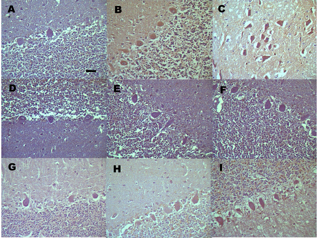
This histopathologic case-control study was designed to characterize the dynamic changes in protein expression of nuclear factor-kappa B (NF-kappaB)/p65 subunit, macrophage inflammatory protein-2 (MIP-2), and matrix metalloproteinase-9 (MMP-9) in postmortem brains of patients with and without intracerebral hemorrhage (ICH). Thirty-six human brains from patients with ICH and six control brains were included in this study. We found that expression levels of NF-kappaB/p65, MIP-2, and MMP-9 were each upregulated on the injured side of the hippocampus at times ranging from 2h to 5days post-ICH. Interestingly, the expression of all three markers was also upregulated on the uninjured side of the hippocampus and in the cerebellum, although to a lesser extent. These data suggest that inflammation occurs early and persists for several days after ICH in humans and could be involved in the progression of ICH-induced secondary brain damage.
Copyright 2010 Elsevier B.V. All rights reserved.
Figures


References
-
- Abilleira S, Montaner J, Molina CA, Monasterio J, Castillo J, Alvarez-Sabin J. Matrix metalloproteinase-9 concentration after spontaneous intracerebral hemorrhage. J Neurosurg. 2003;99:65–70. - PubMed
-
- Alvarez-Sabin J, Delgado P, Abilleira S, Molina CA, Arenillas J, Ribo M, Santamarina E, Quintana M, Monasterio J, Montaner J. Temporal profile of matrix metalloproteinases and their inhibitors after spontaneous intracerebral hemorrhage: relationship to clinical and radiological outcome. Stroke. 2004;35:1316–1322. - PubMed
-
- Bell MD, Taub DD, Kunkel SJ, Strieter RM, Foley R, Gauldie J, Perry VH. Recombinant human adenovirus with rat MIP-2 gene insertion causes prolonged PMN recruitment to the murine brain. Eur J Neurosci. 1996;8:1803–1811. - PubMed
-
- Castellazzi M, Tamborino C, De Santis G, Garofano F, Lupato A, Ramponi V, Trentini A, Casetta I, Bellini T, Fainardi E. Timing of serum active MMP-9 and MMP-2 levels in acute and subacute phases after spontaneous intracerebral hemorrhage. Acta Neurochir Suppl. 2010;106:137–140. - PubMed
-
- Chen Y, Fan Y, Poon KY, Achrol AS, Lawton MT, Zhu Y, McCulloch CE, Hashimoto T, Lee C, Barbaro NM, Bollen AW, Yang GY, Young WL. MMP-9 expression is associated with leukocytic but not endothelial markers in brain arteriovenous malformations. Front Biosci. 2006;11:3121–3128. - PubMed
Publication types
MeSH terms
Substances
Grants and funding
LinkOut - more resources
Full Text Sources
Medical
Miscellaneous
