

Development and validation of a heart atlas to study cardiac exposure to radiation following treatment for breast cancer
- PMID: 20421148
- PMCID: PMC2937165
- DOI: 10.1016/j.ijrobp.2009.10.058
Development and validation of a heart atlas to study cardiac exposure to radiation following treatment for breast cancer
Abstract
Purpose: Cardiac toxicity is an important sequela of breast radiotherapy. However, the relationship between dose to cardiac structures and subsequent toxicity has not been well defined, partially due to variations in substructure delineation, which can lead to inconsistent dose reporting and the failure to detect potential correlations. Here we have developed a heart atlas and evaluated its effect on contour accuracy and concordance.
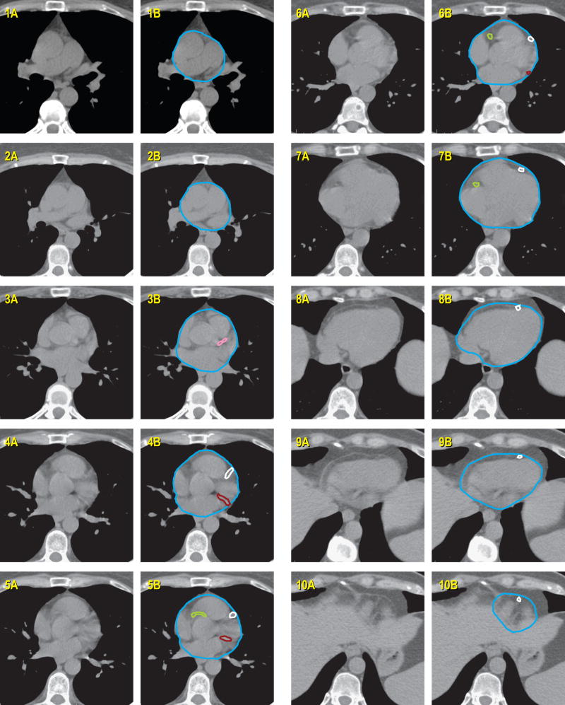
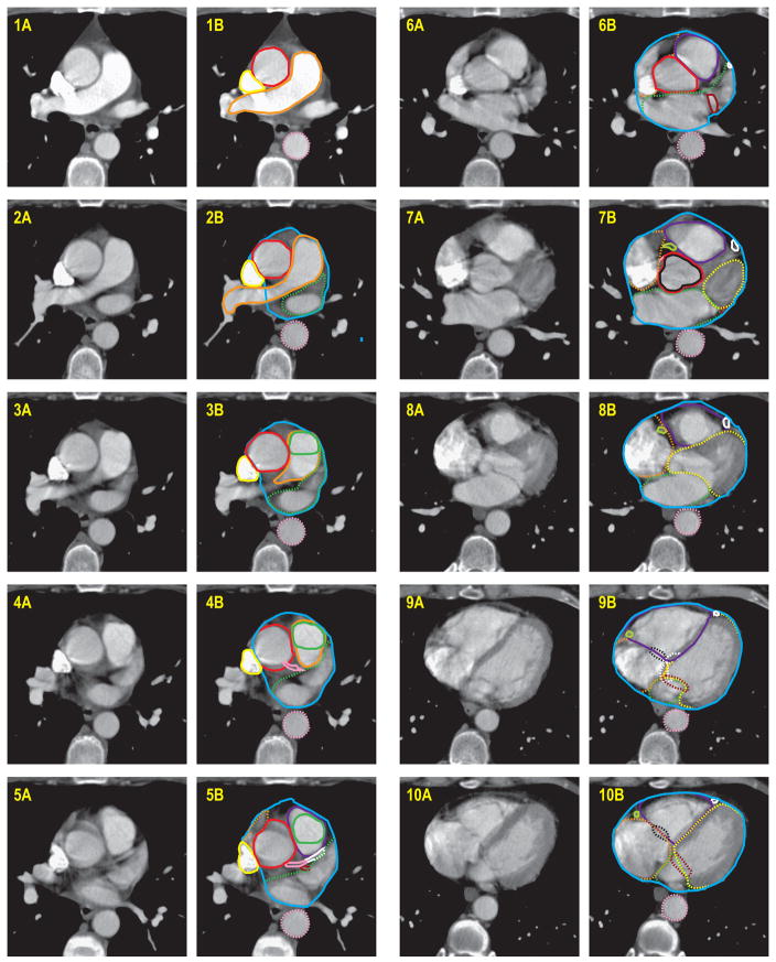
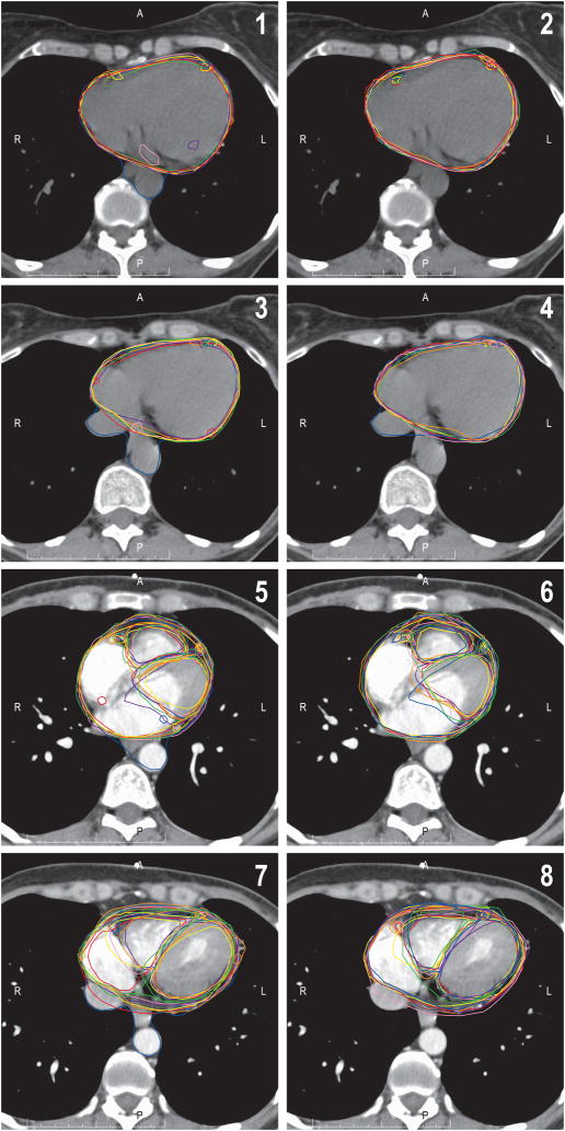
Methods and materials: A detailed cardiac computed tomography scan atlas was developed jointly by cardiology, cardiac radiology, and radiation oncology. Seven radiation oncologists were recruited to delineate the whole heart, left main and left anterior descending interventricular branches, and right coronary arteries on four cases before and after studying the atlas. Contour accuracy was assessed by percent overlap with gold standard atlas volumes. The concordance index was also calculated. Standard radiation fields were applied. Doses to observer-contoured cardiac structures were calculated and compared with gold standard contour doses. Pre- and post-atlas values were analyzed using a paired t test.
Results: The cardiac atlas significantly improved contour accuracy and concordance. Percent overlap and concordance index of observer-contoured cardiac and gold standard volumes were 2.3-fold improved for all structures (p < 0.002). After application of the atlas, reported mean doses to the whole heart, left main artery, left anterior descending interventricular branch, and right coronary artery were within 0.1, 0.9, 2.6, and 0.6 Gy, respectively, of gold standard doses.
Conclusions: This validated University of Michigan cardiac atlas may serve as a useful tool in future studies assessing cardiac toxicity and in clinical trials which include dose volume constraints to the heart.
Copyright © 2011 Elsevier Inc. All rights reserved.
Conflict of interest statement
No actual or potential conflicts of interest exist.
Figures

References
-
- Hooning MJ, Botma A, Aleman BM, et al. Long-term risk of cardiovascular disease in 10-year survivors of breast cancer. J Natl Cancer Inst. 2007;99:365–375. - PubMed
-
- Harris EE, Correa C, Hwang WT, et al. Late cardiac mortality and morbidity in early-stage breast cancer patients after breast-conservation treatment. J Clin Oncol. 2006;24:4100–4106. - PubMed
-
- Jagsi R, Griffith KA, Koelling T, et al. Rates of myocardial infarction and coronary artery disease and risk factors in patients treated with radiation therapy for early-stage breast cancer. Cancer. 2007;109:650–657. - PubMed
-
- Clarke M, Collins R, Darby S, et al. Effects of radiotherapy and of differences in the extent of surgery for early breast cancer on local recurrence and 15-year survival: an overview of the randomised trials. Lancet. 2005;366:2087–2106. - PubMed
-
- Heidenreich PA, Schnittger I, Strauss HW, et al. Screening for coronary artery disease after mediastinal irradiation for Hodgkin's disease. J Clin Oncol. 2007;25:43–49. - PubMed
Publication types
MeSH terms
Substances
Grants and funding
LinkOut - more resources
Full Text Sources
Other Literature Sources
Medical
