. 2010 Sep;38(16):5443-55.
doi: 10.1093/nar/gkq284. Epub 2010 Apr 26.

BpaB, a novel protein encoded by the Lyme disease spirochete's cp32 prophages, binds to erp Operator 2 DNA

Affiliations

BpaB, a novel protein encoded by the Lyme disease spirochete's cp32 prophages, binds to erp Operator 2 DNA

Logan H Burns et al. Nucleic Acids Res. 2010 Sep.

Abstract

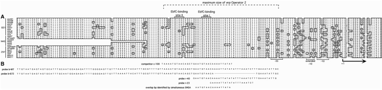
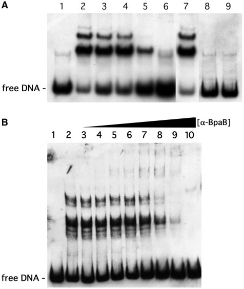
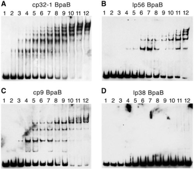
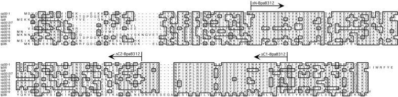
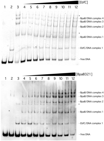
Borrelia burgdorferi produces Erp outer surface proteins throughout mammalian infection, but represses their synthesis during colonization of vector ticks. A DNA region 5' of the start of erp transcription, Operator 2, was previously shown to be essential for regulation of expression. We now report identification and characterization of a novel erp Operator 2-binding protein, which we named BpaB. erp operons are located on episomal cp32 prophages, and a single bacterium may contain as many as 10 different cp32s. Each cp32 family member encodes a unique BpaB protein, yet the three tested cp32-encoded BpaB alleles all bound to the same DNA sequence. A 20-bp region of erp Operator 2 was determined to be essential for BpaB binding, and initial protein binding to that site was required for binding of additional BpaB molecules. A 36-residue region near the BpaB carboxy terminus was found to be essential for high-affinity DNA-binding. BpaB competed for binding to erp Operator 2 with a second B. burgdorferi DNA-binding protein, EbfC. Thus, cellular levels of free BpaB and EbfC could potentially control erp transcription levels.


Figures

Figure 1.
Figure 1.
(A) DNA sequences 5′ of all known erp loci of B. burgdorferi strains B31, N40 and 297, indicating transcriptional promoter motifs, start of transcription (+1), the maximal borders of Operator 2 and two EbfC-binding sequences. Identical nucleotides found in the majority of known erp loci are boxed and shaded. For some erp loci of strain N40, only parts of some 5′ sequences are known, as indicated by blanks on this figure. These three strains, plus B. burgdorferi strain BL206 and Sh-2-82, are the only Lyme disease spirochetes for which the complete erp locus sequences have been published. The erp locus sequences of strain BL206 are identical to those of strain B31, and those of Sh-2-82 are identical to the erp loci of strain 297, and so were omitted from this figure (our unpublished data and references 18, 40, 43 and 44). (B) DNA sequences of biotin- and radiolabeled DNA probes used in EMSA studies. Base pairs present in the wild-type probe (b-WT) but absent from probe b-672 are indicated by a line. The DNA sequence identified as including the initial BpaB-binding site through studies illustrated in Figure 5 are shown on the lower line. All EMSA probes and competitors were double-stranded DNAs; for simplicity, only one strand (5′ to 3′) is illustrated.
Figure 2.
Figure 2.
Demonstration that BpaB binds specifically to erp Operator 2 DNA. (A) EMSA with labeled erp probe b-WT and purified recombinant protein based on the sequence of bpaB from B. burgdorferi strain B31 cp32-1 (BpaB321). Lane 1 lacked added BpaB321, while 0.12 µM recombinant protein was added to the other lanes. (Lanes 2–4) Pre-incubation of DNA and protein at room temperature, 4°C and 37°C, respectively. (Lanes 5–7) DNA and protein mixtures were preincubated with 100-fold excesses of unlabeled competitor DNAs C-100, unlabeled-WT, or flaB, respectively. (Lane 8) BpaB321 was heated in a boiling water bath for 10 min prior to mixture with DNA. (Lane 9) 10% SDS, final concentration, was added to the DNA–protein mixture prior to EMSA. (B) Supershift EMSA using BpaB-directed antibodies. Lane 1 contained only DNA, lane 2 contained DNA plus 0.19 µM BpaB321, and lanes 3–10 contained the same concentrations of DNA and BpaB321 plus increasing concentrations of anti-BpaB321 polyclonal antiserum.
Figure 3.
Figure 3.
EMSA using labeled probe b-WT and increasing concentrations of recombinant BpaB proteins based on sequences of B31 plasmids cp32-1, lp56, cp9-1 and lp38. For all EMSAs, lane 1 contained DNA without protein. (A) Lanes 2–12 contained in addition 0.033, 0.066, 0.099, 0.13, 0.17, 0.20, 0.23, 0.26, 0.30, 0.33 and 0.40 µM recombinant BpaB321 protein, respectively. (B) Lanes 2–12 contained 0.0048, 0.012, 0.024, 0.048, 0.097, 0.15, 0.19, 0.24, 0.29, 0.34 and 0.39 µM recombinant BpaB56 protein, respectively. (C) Lanes 2–12 contained 0.084, 0.17, 0.25, 0.34, 0.42, 0.50, 0.59, 0.67, 0.75, 0.84 and 1.00 µM recombinant BpaB9 protein, respectively. (D) Lanes 2–12 contained 0.069, 0.14, 0.21, 0.28, 0.35, 0.42, 0.48, 0.55, 0.62, 0.69 and 0.83 µM recombinant BpaB38 protein, respectively. Note that some of the higher-order DNA–protein complexes do not always resolve crisply, and appear faint or as smears in EMSAs [e.g. the upper EMSA bands in lane 12 of (C)].
Figure 4.
Figure 4.
Alignments of the predicted amino acid sequences of BpaB proteins encoded by B. burgdorferi B31 cp32 family members, and by the strain B31 plasmid lp38. Identical residues found in the majority of proteins are boxed and shaded. Gaps introduced to maximize homology are indicated by dashes. The amino terminus of protein ΔN-BpaB321 and the carboxy-termini of proteins ΔC1-BpaB321 and ΔC2-BpaB321 are indicated.
Figure 5.
Figure 5.
Identification of the high-affinity BpaB-binding site in erp Operator 2, by use of simultaneous titration of multiple probes with increasing concentrations of BpaB321. In this method, several DNAs that overlap in sequence were simultaneously incubated with protein, then subjected to EMSA. These DNAs which contain the high-affinity protein binding site were preferentially bound by lower concentrations of protein, and signals corresponding with those free DNA disappeared soonest. (A) Sequences of labeled DNA probes used in simultaneous EMSAs. Asterisks indicate DNAs preferentially bound by BpaB. The –35 sequence of the erpAB promoter and the two consensus EbfC-binding sites are indicated by underlining. The upper seven nested DNAs were anchored 51 bases downstream of the erpAB transcription start site, and EMSA data are illustrated in (B). The lower seven nested DNAs were anchored at bp –174 relative to the start of erpAB transcription, and EMSA data are illustrated in (C). The sequence contained in all DNAs that preferentially bound BpaB but absent from lower-affinity DNAs is indicated by a thick horizontal bar. (B) Simultaneous EMSA of recombinant BpaB321 with the upper seven DNAs of (A). Lane 1 contains only the DNAs. Lanes 2–12 contain DNAs plus increasing concentrations of recombinant BpaB321. Quantification of signal strengths indicated that BpaB bound preferentially to the largest DNA (marked with an arrowhead): in lanes 9, 10 and 12, the proportion of free DNA for this probe was 21, 0.10 and 0% of lane 1, respectively, while that of the next largest probe was 52, 13 and 10% of lane 1, respectively. (C) Simultaneous EMSA of recombinant BpaB321 with the lower seven DNAs of (A). Lane 1 contains only the DNAs. Lanes 2–12 contain DNAs plus increasing concentrations of recombinant BpaB321. Quantification of signal strengths indicated that BpaB bound preferentially to the five largest DNAs (marked with arrowheads): in lanes 7, 8 and 9, the proportion of free DNA for the largest probe was 16, 10 and 0% of lane 1, respectively, while that of the smallest probe was 75, 55 and 43% of lane 1, respectively.
Figure 6.
Figure 6.
Binding of BpaB changes DNA conformation. (A) Sequence of the 5′ end of the erpAB1 locus (top line) and five biotinylated 150-bp fragments of that DNA (probes 1 to 5). Arrowhead under the panel indicates the center of probe 3. (B) Comparisons of mobilities of free probes 1–5 and those probes bound by BpaB321. Numbers above each lane indicate the labeled probe being analyzed. Note that free probe 5 migrates the slowest, indicative of a naturally occurring bend near the center of that DNA (22). Binding of BpaB321 resulted in probe 3 migrating the slowest, indicating an induced change in DNA conformation.
Figure 7.
Figure 7.
Confirmation of the initial BpaB-binding site in erp Operator 2 (A) Simultaneous EMSA using wild-type recombinant BpaB321 protein and both biotin-labeled wild-type erpAB1 probe b-WT and b-672. Probe b-672 is identical to b-WT except for deletion of 20 bp (Figure 1). Lane 1 contained only DNA. Lanes 2–12 contained BpaB321 at concentrations of 0.086, 0.18, 0.26, 0.35, 0.44, 0.53, 0.61, 0.70, 0.79, 0.86 and 1.1 µM, respectively. (B) EMSA using the radioactively labeled 40 bp probe r-40 (Figure 1). Lane 1 contained only DNA. Lanes 2–20 contained BpaB321 at concentrations of 0.40, 0.80, 1.0, 1.2, 1.5, 2.0, 2.5, 3.0, 3.5, 4.0, 5.0, 7.0, 10, 15, 20, 25, 30, 40 and 50 µM, respectively. (C) EMSA using the radioactively labeled 23-bp probe r-23, which encompasses the 20-bp sequence present in b-WT but absent from b-672 (see Figure 1). Lane 1 contained only DNA. Lanes 2–17 contained BpaB321 at concentrations of 0.40, 0.80, 1.0, 1.2, 1.5, 2.0, 2.5, 3.0, 3.5, 4.0, 5.0, 7.0, 10, 15, 20 and 30 µM, respectively.
Figure 8.
Figure 8.
EMSAs using wild-type erpAB1 DNA and truncated BpaB321 proteins. For all panels, lane 1 contained DNA without protein. (A) Lanes 2–12 contained 0.013, 0.026, 0.039, 0.052, 0.065, 0.078, 0.090, 0.10, 0.12, 0.13 and 0.16 µM recombinant ΔN-BpaB321, respectively. (B) Lanes 2–12 contained 0.078, 0.016, 0.023, 0.031, 0.039, 0.047, 0.055, 0.062, 0.070, 0.078 and 0.094 µM recombinant ΔC1-BpaB321 respectively. (C) Lanes 2–12 contained 0.0075, 0.015, 0.023, 0.030, 0.038, 0.045, 0.053, 0.060, 0.068, 0.075 and 0.090 µM recombinant ΔC2-BpaB321, respectively.
Figure 9.
Figure 9.
EbfC and BpaB compete for binding to erp Operator 2. (A) EMSAs in which the concentration of BpaB321 was held constant while increasing concentrations of EbfC were added. (Lane 1) Labeled probe b-WT alone. (Lanes 2 and 3) Probe b-WT preincubated with 2.05 and 2.89 µM recombinant EbfC or BpaB321, respectively. (Lanes 4–12) Probe b-WT preincubated with 0.97 µM recombinant BpaB321, plus EbfC at concentrations of 0.27, 0.51, 1.03, 1.54, 2.05, 2.56, 3.08, 3.59 and 4.10 µM, respectively. Compositions of EMSA bands are indicated to the right of the panel. Asterisks indicate nonspecific DNA binding by EbfC observed at high concentrations of that protein (our unpublished data). Ratios of free DNA to BpaB–DNA complex 1 in lanes 4 through 12 were 3.9 : 1, 3.5 : 1, 2.7 : 1, 2.1 : 1, 1.4 : 1, 1.1 : 1, 1.1 : 1, 0.86 : 1 and 0.57 : 1, respectively, with the BpaB–DNA complex signals remaining constant, indicating that EbfC preferentially bound to free DNA. Quantification of signal intensities indicated that the proportion of EbfC–DNA complexes steadily increased with addition of EbfC protein, and that total of signal strengths remained constant across the EMSA gel. (B) EMSAs in which the concentration of EbfC was held constant while increasing concentrations of BpaB321 were added. (Lane 1) Labeled probe b-WT alone. (Lanes 2 and 3) Probe b-WT pre-incubated with 0.2 and 0.3 µM recombinant EbfC or BpaB321, respectively. (Lanes 4–12) Probe b-WT preincubated with 0.3 µM recombinant BpaB321, plus EbfC at concentrations of 0.2, 0.3, 0.4, 0.5, 0.6, 0.7, 0.8, 0.9 and 1 µM, respectively. Compositions of EMSA bands are indicated to the right of the panel. The asterisk indicates nonspecific DNA binding by EbfC observed at high concentrations of that protein (our unpublished data). Quantification of signal strengths indicated that proportions of free DNA and DNA bound by EbfC decreased as additional BpaB was added, while proportions of BpaB–DNA complexes increased.

Similar articles

Cited by

References

    1. Stanek G, Strle F. Lyme borreliosis. Lancet. 2003;362:1639–1647. - PubMed
    1. Steere AC. Lyme disease. New Engl. J. Med. 2001;345:115–125. - PubMed
    1. Stevenson B, Zückert WR, Akins DR. In: The Spirochetes: Molecular and Cellular Biology. Saier MH, García-Lara J, editors. Oxford: Horizon Press; 2001. pp. 87–100.
    1. Stevenson B, Bykowski T, Cooley AE, Babb K, Miller JC, Woodman ME, von Lackum K, Riley SP. In: Molecular Biology of Spirochetes. Cabello FC, Godfrey HP, Hulinska D, editors. Amsterdam: IOS Press; 2006. pp. 354–372.
    1. Brissette CA, Verma A, Bowman A, Cooley AE, Stevenson B. The Borrelia burgdorferi outer-surface protein ErpX binds mammalian laminin. Microbiology. 2009;155:863–872. - PMC - PubMed

Publication types

MeSH terms