. 2010 Jun 15;588(Pt 12):2255-67.
doi: 10.1113/jphysiol.2009.186643. Epub 2010 Apr 26.

Receptor-promoted exocytosis of airway epithelial mucin granules containing a spectrum of adenine nucleotides

Affiliations

Receptor-promoted exocytosis of airway epithelial mucin granules containing a spectrum of adenine nucleotides

Silvia M Kreda et al. J Physiol. .

Abstract

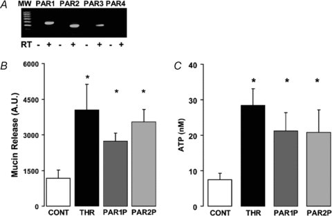
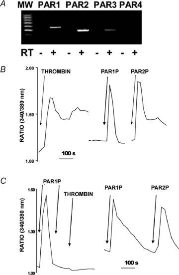
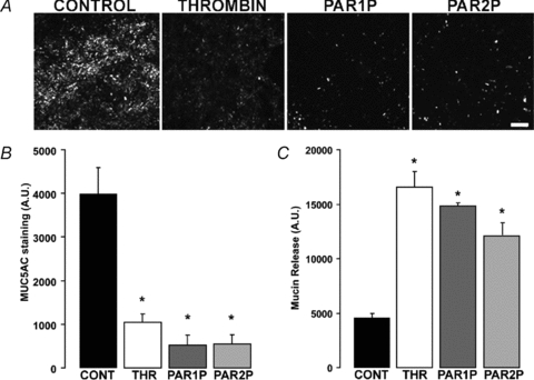
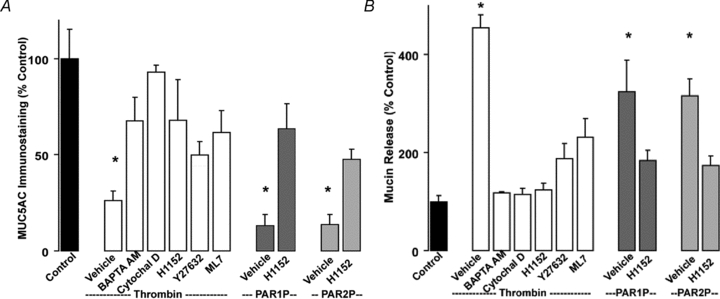
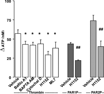
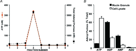
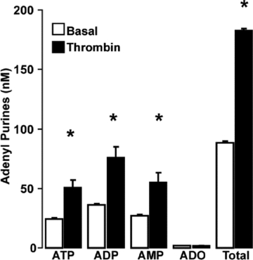
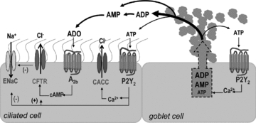
Purinergic regulation of airway innate defence activities is in part achieved by the release of nucleotides from epithelial cells. However, the mechanisms of airway epithelial nucleotide release are poorly understood. We have previously demonstrated that ATP is released from ionomycin-stimulated airway epithelial goblet cells coordinately with mucin exocytosis, suggesting that ATP is released as a co-cargo molecule from mucin-containing granules. We now demonstrate that protease-activated-receptor (PAR) agonists also stimulate the simultaneous release of mucins and ATP from airway epithelial cells. PAR-mediated mucin and ATP release were dependent on intracellular Ca(2+) and actin cytoskeleton reorganization since BAPTA AM, cytochalasin D, and inhibitors of Rho and myosin light chain kinases blocked both responses. To test the hypothesis that ATP is co-released with mucin from mucin granules, we measured the nucleotide composition of isolated mucin granules purified based on their MUC5AC and VAMP-8 content by density gradients. Mucin granules contained ATP, but the levels of ADP and AMP within granules exceeded by nearly 10-fold that of ATP. Consistent with this finding, apical secretions from PAR-stimulated cells contained relatively high levels of ADP/AMP, which could not be accounted for solely based on ATP release and hydrolysis. Thus, mucin granules contribute to ATP release and also are a source of extracellular ADP and AMP. Direct release of ADP/AMP from mucin granules is likely to provide a major source of airway surface adenosine to signal in a paracrine faction ciliated cell A(2b) receptors to activate ion/water secretion and appropriately hydrate goblet cell-released mucins.


Figures

Figure 1
Figure 1. PAR agonists stimulate mucin and ATP release from WD-HBE cells
A, RT-PCR analysis indicating that PAR1, PAR2 and PAR3 (but not PAR4) transcripts were amplified in WD-HBE cells; RT, reverse transcriptase. B and C, WD-HBE cultures were incubated basolaterally with vehicle, 50 nm thrombin, 100 μm PAR1P, or 100 μm PAR2P for 5 min at 37°C. The apical bath was analysed for mucin content by immuno-slot blot (B) and ATP content by the luciferin–luciferase assay (C). Experiments were performed in quadruplicate with cultures from three different donors. The results are expressed as the mean ±s.e.m. (*P < 0.01).
Figure 2
Figure 2. Calu-3 express PARs
A, RT-PCR analysis indicating that PAR1, PAR2 and PAR3 (but not PAR4) transcripts were expressed in Calu-3 cells. B, intracellular calcium mobilization was assessed in independent cultures of Fura-2-loaded Calu-3 cells exposed to 50 nm thrombin, 100 μm PAR1P, or 100 μm PAR2P. C, left, fura-2 loaded cells were challenged with 100 μm PAR1AP and, after the Ca2+ signal relaxed, a second dose of 100 μm PAR1AP was added followed by 50 nm thrombin (left tracing); right, Calu-3 cells were exposed to 100 μm PAR1AP and then to 100 μm PARA2P. The tracings are representative of three independent experiments performed in duplicate.
Figure 3
Figure 3. PAR agonists stimulate mucin release from Calu-3 cells
Calu-3 cells were challenged with vehicle, 50 nm thrombin, 100 μm PAR1P, or 100 μm PAR2P for 5 min at 37°C. A, mucin granule content was determined by immunostaining with a MUC5AC antibody followed by confocal microscopy analysis (bar = 100 μm). B, quantification of MUC5AC immunostaining in Calu-3 cultures. C, mucin release in the lumenal bath was assessed by slot blot as in Fig. 1. The results of a representative experiment are illustrated, and the data are expressed in arbitrary units and are the mean ±s.e.m. (n= 4; *P < 0.01). Similar results were obtained in three independent experiments.
Figure 4
Figure 4. PAR-stimulated mucin release is Ca2+ and cytoskeleton dependent
Calu-3 cells were pre-incubated for 30 min at 37°C with vehicle, 10 μm BAPTA AM, 5 μm cytochalasin D, 100 nm H1152, 10 μm Y27632, or 1 μm ML7. Cells were challenged with vehicle, 50 nm thrombin, 100 μm PAR1P, or 100 μm PAR2P for 5 min at 37°C. A, mucin granule content was quantified by immunostaining as in Fig. 3. B, mucin release in the apical bath was assessed by slot blot as in Fig. 1. Experiments were performed three times, each condition in quadruplicate. The results of a representative experiment are illustrated and data are expressed as percentage of control (mean ±s.e.m.; *P < 0.01 vs. control).
Figure 5
Figure 5. PAR-stimulated ATP release involves a vesicular, Ca2+- and cytoskeleton- dependent mechanism
Calu-3 cells were pre-incubated with inhibitors as in Fig. 4, or with 4 μm Bafilomycin A1 for 30 min at 37°C. Mucosal ATP release following the addition of the indicated PAR agonists (5 min at 37°C) was assessed using the luciferin–luciferase assay in the presence of blockers of ecto-nucleotidases. Experiments were performed in quadruplicate with three independent cultures. The results of a representative experiment are illustrated, and the data are expressed as the difference between PAR agonist and basal values (basal ATP values, 15 ± 5 nm) (mean ±s.e.m.; *P < 0.01 compared to thrombin stimulation; ##P < 0.01 compared to PAR1P and PAR2P stimulation).
Figure 6
Figure 6. PAR agonists stimulate secretion of quinacrine-labelled granules
Calu-3 cell mucin granules were loaded with quinacrine (10 μm, 20 min at 37°C). Cells were mounted in a confocal microscope and real-time images of the DIC/Nomarski illumination (grey) and fluorescence (green) channels acquired every 30 s (see Methods). Cells were challenged with vehicle (control), 50 nm thrombin, 100 μm PAR1P, or 100 μm PAR2P. A, overlay of the DIC and fluorescence confocal images of quinacrine-labelled Calu-3 cells in control conditions; bar = 10 μm. B, representation of the change in fluorescence intensity associated with 1 μm granules after 5 min incubation with vehicle or PAR agonists (n= 3; mean ±s.e.m.; *P < 0.01).
Figure 7
Figure 7. Isolation of mucin granules from Calu-3 cells
Mucin granules were isolated from Calu-3 cells using two consecutive Percoll gradients as described in Methods; the results of a representative isolation experiment are illustrated. A, confocal microscopy image (DIC/fluorescence channel overlay) of an isolated mucin granule from Calu-3 cultures labelled with quinacrine. B, image of the immuno-slot blot for MUC5AC representing the first eight fractions of the second gradient. C, the profile of organelle distribution in the second gradient fractions was assessed by immuno-slot blot using specific antibodies that recognize the indicated cellular markers. The quantification of the densitometry data for each organelle marker is expressed as a percentage of the total lysate content. D, localization of VAMP-8 and MUC5AC was assessed by immunostaining under resting (control; left panels) or thrombin-stimulated (50 nm, 5 min at 37°C; right panels) conditions in Calu-3 cells; bar = 10 μm.
Figure 8
Figure 8. Isolated mucin granules contain ATP and other nucleotides
A, quantification of the amounts of ATP and total adenyl purine in each of the fractions collected in the second gradient was performed by etheno-derivatization and HPLC analysis. Data are expressed as the concentration of ATP (left axis) and total adenine-containing species (right axis); note that left and right axes represent different concentration ranges. B, quantification of the content of adenyl purine species (ATP, ADP, AMP and adenosine) in the total cell lysate and isolated mucin granule fraction (i.e. fraction 4) was performed by etheno-derivatization and HPLC analysis. Data are the average of four independent granule isolations and represent the percentage distribution of each species with respect to the total adenyl purine content in the fraction (mean ±s.e.m.). Note, the total adenyl purine mass in the mucin granule fraction represented <5% of the cell lysate content.
Figure 9
Figure 9. Nucleotide composition of Calu-3 cell secretions
Calu-3 cells were stimulated with thrombin (50 nm, 5 min at 37°C) and the apical bath was collected and analysed for adenyl purines as above. The results of a representative experiment are illustrated, and data are expressed as the mean ±s.e.m.; *P < 0.01 compared to non-stimulated (basal) levels. Similar results were obtained in two independent experiments performed in quadruplicate.
Figure 10
Figure 10. Model of adenyl nucleotide regulation in ASL
The schematic diagram represents a ciliated and a goblet cell of the airway surface epithelium. Mucin exocytosis from goblet cells is accompanied by release of adenyl nucleotides present in mucin granules as co-cargo molecules. ADP is the prevalent species followed by AMP and ATP. In ASL, ADP and AMP (and ATP) are rapidly metabolized by ecto-nucleotidases into adenosine. Adenyl purines have autocrine and paracrine regulatory activities on epithelial cells. For example, adenosine stimulates the A2b receptor on ciliated cells. CFTR, which is expressed in ciliated cells, is activated by A2b receptor-promoted cAMP formation (and PKA activation, not shown). Thus, chloride secretion is increased and sodium absorption is reduced (by CFTR-mediated inhibition of ENaC), which generates the driving gradient for water secretion necessary to disperse newly secreted mucins into the ASL. ATP released from mucin granules stimulates P2Y2 receptors on goblet cells for further mucin secretion, and on ciliated cells resulting in activation of TMEM16A or CACC (calcium activated chloride channel), activation of CFTR (via PKC), and inhibition of ENaC.

Similar articles

Cited by

References

    1. Bankston LA, Guidotti G. Characterization of ATP transport into chromaffin granule ghosts. Synergy of ATP and serotonin accumulation in chromaffin granule ghosts. J Biol Chem. 1996;271:17132–17138. - PubMed
    1. Boucher RC. Airway surface dehydration in cystic fibrosis: pathogenesis and therapy. Annu Rev Med. 2007;58:157–170. - PubMed
    1. Boudreault F, Grygorczyk R. Cell swelling-induced ATP release is tightly dependent on intracellular calcium elevations. J Physiol. 2004;561:499–513. - PMC - PubMed
    1. Davis CW, Dickey BF. Regulated airway goblet cell mucin secretion. Annu Rev Physiol. 2008;70:487–512. - PubMed
    1. Davis CW, Dowell ML, Lethem MI, Van Scott M. Goblet cell degranulation in isolated canine tracheal epithelium: Response to exogenous ATP, ADP, and adenosine. Am J Physiol Cell Physiol. 1992;262:C1313–C1323. - PubMed

Publication types

MeSH terms

LinkOut - more resources