

Peripheral chemoreceptors determine the respiratory sensitivity of central chemoreceptors to CO(2)
- PMID: 20421288
- PMCID: PMC2915520
- DOI: 10.1113/jphysiol.2010.187211
Peripheral chemoreceptors determine the respiratory sensitivity of central chemoreceptors to CO(2)
Abstract
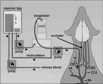
We assessed the contribution of carotid body chemoreceptors to the ventilatory response to specific CNS hypercapnia in eight unanaesthetized, awake dogs. We denervated one carotid body (CB) and used extracorporeal blood perfusion of the reversibly isolated remaining CB to maintain normal CB blood gases (normoxic, normocapnic perfusate), to inhibit (hyperoxic, hypocapnic perfusate) or to stimulate (hypoxic, normocapnic perfusate) the CB chemoreflex, while the systemic circulation, and therefore the CNS and central chemoreceptors, were exposed consecutively to four progressive levels of systemic arterial hypercapnia via increased fractional inspired CO(2) for 7 min at each level. Neither unilateral CB denervation nor CB perfusion, per se, affected breathing. Relative to CB control conditions (normoxic, normocapnic perfusion), we found that CB chemoreflex inhibition decreased the slope of the ventilatory response to CNS hypercapnia in all dogs to an average of 19% of control values (range 0-38%; n = 6), whereas CB chemoreflex stimulation increased the slope of the ventilatory response to CNS hypercapnia in all dogs to an average of 223% of control values (range 204-235%; n = 4). We conclude that the gain of the CNS CO(2)/H(+) chemoreceptors in dogs is critically dependent on CB afferent activity and that CNS-CB interaction results in hyperadditive ventilatory responses to central hypercapnia.
Figures
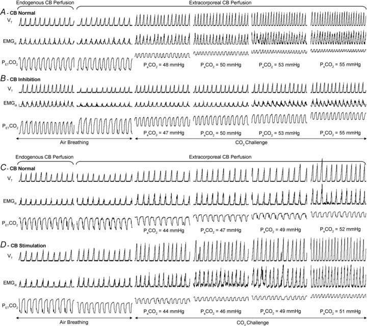
 (segments 3–6). Each segment represents 1 min of recorded data. Abbreviations: EMGdi, moving-time-averaged electromyogram of the costal diaphragm;
  , arterial
  ;
;  , end-tidal
  ; and VT, tidal volume.
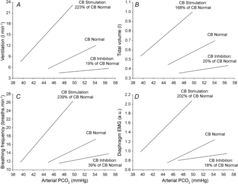
 with a background of extracorporeal carotid body (CB) perfusion with normal (n= 8), inhibitory (n= 6) and stimulatory (n= 4) blood gases. All eight animals are represented in the normal CB central CO2 response mean line. Given the differing number of animals in each group, the mean percentage change in slope relative to CB normal was calculated using only the data for the animals that were represented in the CB inhibition (n= 6) or CB stimulation (n= 4) groups. See main text for details.
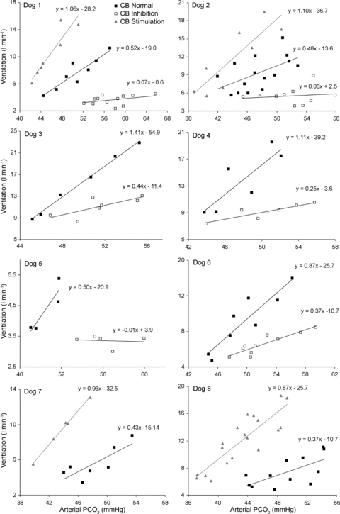
 with a background of extracorporeal CB perfusion with normal, inhibitory and stimulatory perfusate blood gases are shown for each animal in Table 2.
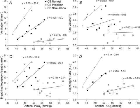
 with a background of extracorporeal carotid body (CB) perfusion with normal (filled squares), inhibitory (open squares) and stimulatory perfusate blood gases (shaded triangles).
Comment in
-
Modulation of the central chemoreflex magnitude by the peripheral chemoreceptors: a hyperadditive effect or are we barking up the wrong tree?J Physiol. 2010 Dec 15;588(Pt 24):4857-8. doi: 10.1113/jphysiol.2010.200246. J Physiol. 2010. PMID: 21173088 Free PMC article. No abstract available.
References
-
- Adams JM, Attinger FM, Attinger EO. Medullary and carotid chemoreceptor interaction for mild stimuli. Pflugers Arch. 1978;374:39–45. - PubMed
-
- Adams JM, Severns ML. Interaction of chemoreceptor effects and its dependence on the intensity of stimuli. J Appl Physiol. 1982;52:602–606. - PubMed
-
- Bellville JW, Whipp BJ, Kaufman RD, Swanson GD, Aqleh KA, Wiberg DM. Central and peripheral chemoreflex loop gain in normal and carotid body-resected subjects. J Appl Physiol. 1979;46:843–853. - PubMed
-
- Bisgard GE, Forster HV, Klein JP. Recovery of peripheral chemoreceptor function after denervation in ponies. J Appl Physiol. 1980;49:964–970. - PubMed
-
- Bisgard GE, Forster HV, Orr JA, Buss DD, Rawlings CA, Rasmussen B. Hypoventilation in ponies after carotid body denervation. J Appl Physiol. 1976;40:184–190. - PubMed
Publication types
MeSH terms
Substances
LinkOut - more resources
Full Text Sources
Other Literature Sources