. 2010 Jun 25;285(26):19842-53.
doi: 10.1074/jbc.M110.129304. Epub 2010 Apr 26.

Xenin-25 potentiates glucose-dependent insulinotropic polypeptide action via a novel cholinergic relay mechanism

Affiliations

Xenin-25 potentiates glucose-dependent insulinotropic polypeptide action via a novel cholinergic relay mechanism

Burton M Wice et al. J Biol Chem. .

Abstract

The intestinal peptides GLP-1 and GIP potentiate glucose-mediated insulin release. Agents that increase GLP-1 action are effective therapies in type 2 diabetes mellitus (T2DM). However, GIP action is blunted in T2DM, and GIP-based therapies have not been developed. Thus, it is important to increase our understanding of the mechanisms of GIP action. We developed mice lacking GIP-producing K cells. Like humans with T2DM, "GIP/DT" animals exhibited a normal insulin secretory response to exogenous GLP-1 but a blunted response to GIP. Pharmacologic doses of xenin-25, another peptide produced by K cells, restored the GIP-mediated insulin secretory response and reduced hyperglycemia in GIP/DT mice. Xenin-25 alone had no effect. Studies with islets, insulin-producing cell lines, and perfused pancreata indicated xenin-25 does not enhance GIP-mediated insulin release by acting directly on the beta-cell. The in vivo effects of xenin-25 to potentiate insulin release were inhibited by atropine sulfate and atropine methyl bromide but not by hexamethonium. Consistent with this, carbachol potentiated GIP-mediated insulin release from in situ perfused pancreata of GIP/DT mice. In vivo, xenin-25 did not activate c-fos expression in the hind brain or paraventricular nucleus of the hypothalamus indicating that central nervous system activation is not required. These data suggest that xenin-25 potentiates GIP-mediated insulin release by activating non-ganglionic cholinergic neurons that innervate the islets, presumably part of an enteric-neuronal-pancreatic pathway. Xenin-25, or molecules that increase acetylcholine receptor signaling in beta-cells, may represent a novel approach to overcome GIP resistance and therefore treat humans with T2DM.


Figures

FIGURE 1.
FIGURE 1.
Xenin-25 potentiates the GIP-mediated reduction in hyperglycemia. Wild-type (WT) or GIP/DT (DT) mice were fasted for 16 h and then given intraperitoneal injections of glucose plus GLP-1 (A), GIP alone (B), xenin-25 alone (Xen; C), or GIP plus xenin-25 (GIP+Xen; D). Blood glucose levels were measured before and at the indicated times after glucose injection. Incremental areas under the curve (E–H) were calculated from data presented in A–D, respectively. All p values compare mice of the same genotype receiving peptide versus BSA without peptide. * and ** represent p values of <0.03 and ≤0.002, respectively.
FIGURE 2.
FIGURE 2.
Xenin-25 potentiates GIP-mediated insulin release in vivo. Male (A) and female (B) mice were treated with BSA, GIP alone (GIP), xenin-25 alone (Xen), or GIP plus xenin-25 (G+X) as described in Fig. 1. Mice receiving saline plus BSA served as fasting controls. Plasma insulin was measured 5 min after the intraperitoneal injection of glucose or saline. p values compare mice with the same genotype receiving glucose plus the indicated peptide(s) versus animals receiving neither glucose nor peptides. * and ** represent p values of <0.05 and 0.005, respectively. The bars indicate p values for mice of the same genotype treated with GIP alone versus GIP plus xenin-25. NS indicates a non-significant p value.
FIGURE 3.
FIGURE 3.
Xenin-25 does not amplify GLP-1 action in mice. A and B, the reduction in hyperglycemia was measured in wild-type (WT) or GIP/DT (DT) mice as described in Fig. 1 except animals received vehicle alone (BSA) or a submaximal dose of GLP-1 (0.1 nmol per mouse) with or without 1 nmol of xenin-25 as indicated. Incremental areas under the curve for A and B are shown in C. D, plasma insulin levels were measured in mice as described in Fig. 2 except animals were injected with either GLP-1 alone or GLP-1 plus xenin-25. E, wild-type (WT) or GIP/DT (DT) mice were treated as described in Fig. 1 except glucose (3 g/kg) was administered at time zero by intragastric gavage rather than intraperitoneal injection. The incremental glucose areas under the curve are shown in F.
FIGURE 4.
FIGURE 4.
Xenin-25 potentiates the GIP-mediated reduction in hyperglycemia in NONcNZO10/LtJ mice. Animals of the indicated age were treated as described in Fig. 1. Areas under the curve for A and B, as well as for 18-week-old mice, are shown in C. * and ** represent p values of <0.05and <0.005, respectively, versus mice receiving BSA alone.
FIGURE 5.
FIGURE 5.
Xenin-25 potentiates GIP-mediated insulin release in NONcNZO10/LtJ mice. Mice of the indicated age were fasted overnight, and plasma insulin was measured before (0) and 15 min after intraperitoneal injections of glucose plus the indicated peptide(s). Control mice were injected with glucose plus vehicle alone (BSA). * and # represent p values of <0.05 and <0.03, respectively, versus mice receiving BSA alone.
FIGURE 6.
FIGURE 6.
Immunoreactive xenin is detectable in mouse plasma following intraperitoneal injection of xenin-25. Wild-type mice were injected with 1 nmol of human gastrin-17 (Gastrin), neurotensin (NT), or xenin-25. Five minutes later, blood was collected and plasma prepared. Immunoreactive xenin was assayed using a commercially available RIA (A) or a custom synthesized ELISA (B). The dotted lines represent the lower limit of detection for both assays and the maximal level detectable by the RIA.
FIGURE 7.
FIGURE 7.
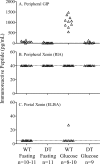
Immunoreactive xenin-25 is not detectable following administration of oral glucose to wild-type (WT) or GIP (DT) mice. Mice were treated as described in Figs. 1 and 2. Blood was collected following a 16-h fast (Fasting) or 15 min after glucose administration (3 g/kg) by intragastric gavage (Glucose). Plasma was prepared and assayed for immunoreactive GIP or xenin using a commercially available ELISA or RIA, respectively. In A and B, blood was collected from the retroorbital sinus of female mice. In C, blood was collected directly from the portal vein of WT mice (4 male and 7 female fasting mice and 6 male and 2 female glucose-treated mice) and assayed for immunoreactive xenin using the custom prepared ELISA.
FIGURE 8.
FIGURE 8.
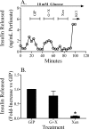
Xenin-25 does not directly stimulate insulin release from the β-cell. Insulin release from islets isolated from wild-type C57BL/6J mice (A; n = 6), GIP/DT mice (B; n = 6) or MIN6 cells (C and D; n = 4) was determined in static incubations. 100 nm GIP, GLP-1, or xenin-25 (Xen) was added to the indicated samples in A and C. In B and D, islets (10 mm glucose) or MIN6 cells (7.5 mm glucose) were incubated with the indicated concentration of GIP in the presence or absence of 10 nm xenin-25. *, p values of <0.02 for groups receiving the indicated peptide versus no peptides (None) at the same concentration of glucose (A and C only).
FIGURE 9.
FIGURE 9.
Xenin-25 does not potentiate GIP-mediated insulin release from the in situ perfused pancreas. The effects of xenin-25 (Xen or X) on GIP (GIP or G)-mediated insulin release were studied using in situ pancreas perfusions in GIP/DT mice (n = 4 perfusions). Glucose was maintained at 10 mm for the duration of the perfusion. Peptides were perfused for 15 min followed by a 15-min washout period. Preliminary perfusions established that 0.3 nm GIP elicited submaximal insulin secretory responses in the presence of 10 mm glucose, which ensured that a xenin-25-mediated increase in insulin release could be detected. The dose of xenin-25 used for the perfusion (3 nm) caused profound activation of xenin-25 signaling in Panc1 cells (see Fig. 10). Representative results from a single mouse are shown in A. The amount of insulin release, normalized to that measured in the presence of GIP alone, is shown for each condition in B. *, p value < 0.03 compared with perfusion with GIP alone.
FIGURE 10.
FIGURE 10.
Incretins, but not xenin-25, increase mitogen-activated protein kinase signaling in MIN6 cells. A, MIN6 cells were treated with the indicated peptide and subjected to Western blot analysis using antibodies specific for activated (P-Erk) or total Erk1/2 (Total Erk). B, Panc-1 exocrine pancreatic cells, which are known to express neurotensin receptors, were treated with the indicated concentration of xenin-25 as described in A. Western blots were probed with antibodies for P-Erk or total Erk.
FIGURE 11.
FIGURE 11.
Xenin-25 potentiation of GIP action is relayed by non-ganglionic, cholinergic neurons. Wild-type (WT) or GIP/DT (DT) mice were treated as described in Fig. 2. As indicated, atropine sulfate (A and B; Atrop), AMB (C), PACAP(6–38) (D; P(6–38)), or hexamethonium chloride (E) was administered to some animals along with the indicated peptide(s). Control mice received vehicle instead of drug. Blood was collected for insulin assays 5 min after injection of glucose. * and ** represent p values of <0.05 and <0.005, respectively. Neither atropine compound exerted a statistically significant effect on WT mice.
FIGURE 12.
FIGURE 12.
Afferent/efferent signaling does not regulate the effects of xenin-25 on GIP action. Following an overnight fast, mice (n = 4 per treatment group) were given an intraperitoneal injection of saline alone (“Saline”), glucose plus GIP (“GIP”), or glucose plus GIP plus xenin-25 (GIP+Xen or G+X). One hour later, brains were harvested for c-fos immunohistochemistry. Representative images of c-fos staining in the DMV and NTS, PVN, or ARC are shown. Brains from insulin injected hypoglycemic mice (“Insulin” or Ins) served as a positive control for induction of c-fos levels in the NTS, PVN, and ARC. A–D, the number of c-fos-positive cells was quantitated by morphometry. *, one-way analysis of variance indicates that variation among column means is significant (p < 0.01), and the Tukey-Kramer multiple comparisons test indicates that the differences in c-fos measured in the presence of insulin, compared with those obtained with saline, GIP plus glucose, or GIP plus glucose plus xenin in the PVN or insulin and GIP plus glucose plus xenin, compared with those obtained with saline or GIP plus glucose in the ARC, are statistically significant (p < 0.05).
FIGURE 13.
FIGURE 13.
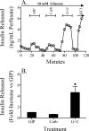
Carbachol potentiates GIP-mediated insulin release from the in situ perfused pancreas. The effects of carbachol (Carb or C) on GIP (GIP or G)-mediated insulin release were studied using in situ pancreas perfusions in GIP/DT mice as described in Fig. 9 (n = 4 perfusions). Preliminary perfusions established that 40 nm carbachol elicited submaximal insulin secretory responses in the presence of 10 mm glucose. Representative results from a single mouse are shown in A. The amount of insulin release, normalized to that measured in the presence of GIP alone, is shown for each condition in B. *, one-way analysis of variance indicates that variation among column means is significant (p = 0.001), and the Tukey-Kramer multiple comparisons test indicates that the differences in insulin release, measured in the presence of GIP plus carbachol compared with those obtained with GIP alone, carbachol alone, or the sum of the values obtained with GIP alone and carbachol alone, are statistically significant (p < 0.05).

Similar articles

Cited by

References

    1. Baggio L. L., Drucker D. J. (2007) Gastroenterology 132, 2131–2157 - PubMed
    1. Kim W., Egan J. M. (2008) Pharmacol. Rev. 60, 470–512 - PMC - PubMed
    1. Brand S. J., Schmidt W. E. (1995) in Textbook of Gastroenterology (Yamada T. ed) pp. 25–71, J. B. Lippincott Co., Philadelphia
    1. Fehmann H., Goke R., Goke B. (1995) Endocrine Rev. 16, 390–410 - PubMed
    1. Walsh J. H. (1994) in Physiology of the Gastrointestinal Tract (Johnson L. R. ed) pp. 1–128, Raven Press, New York

Publication types

MeSH terms

LinkOut - more resources