

Meniscal pathology on MRI increases the risk for both incident and enlarging subchondral bone marrow lesions of the knee: the MOST Study
- PMID: 20421344
- PMCID: PMC2966967
- DOI: 10.1136/ard.2009.121681
Meniscal pathology on MRI increases the risk for both incident and enlarging subchondral bone marrow lesions of the knee: the MOST Study
Abstract
Objectives: To investigate the association between meniscal pathology and incident or enlarging bone marrow lesions (BML) in knee osteoarthritis.
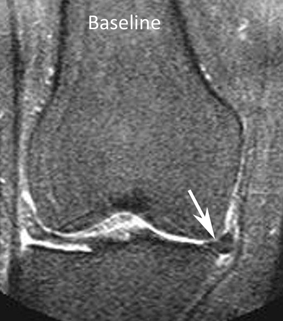
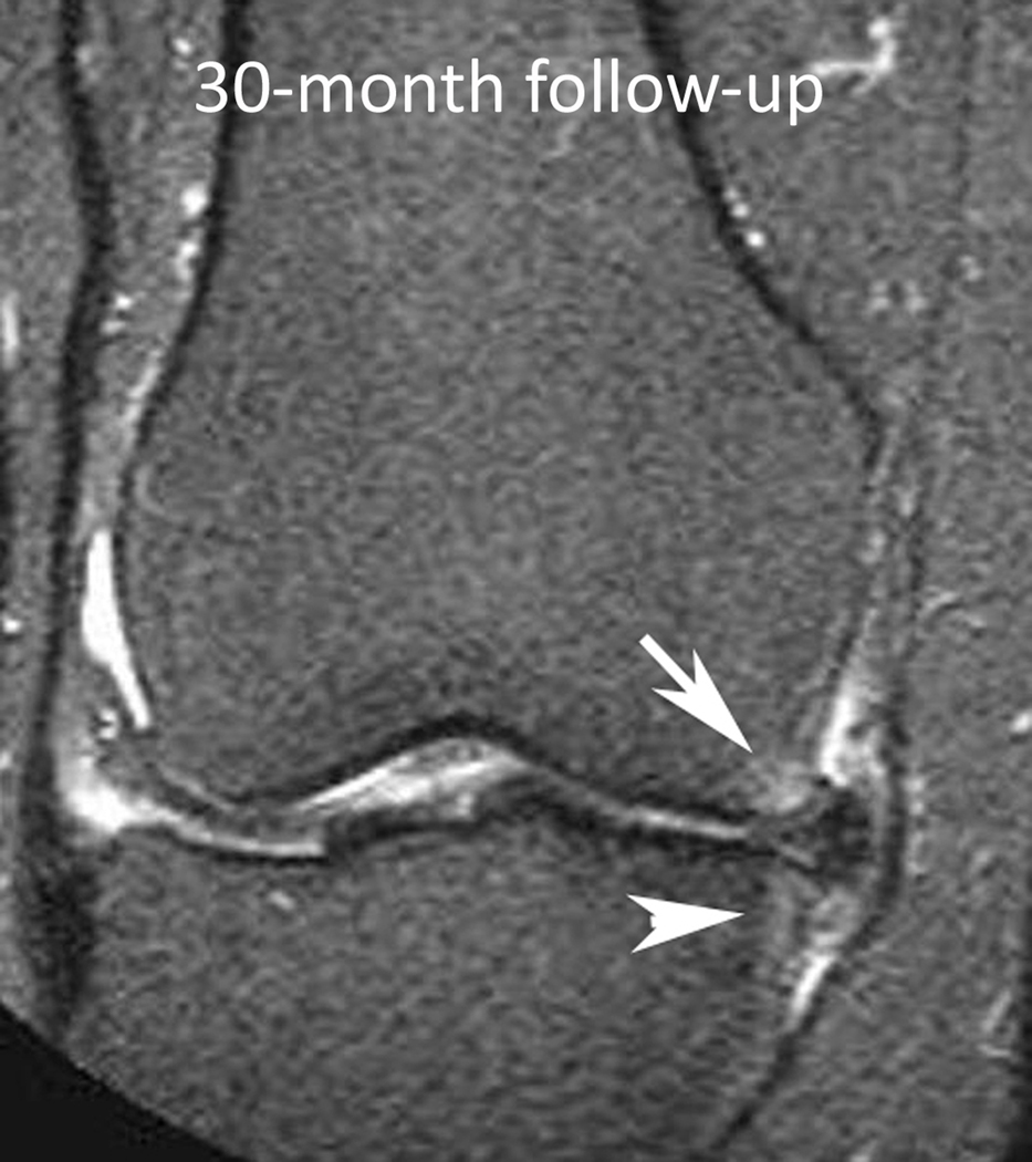
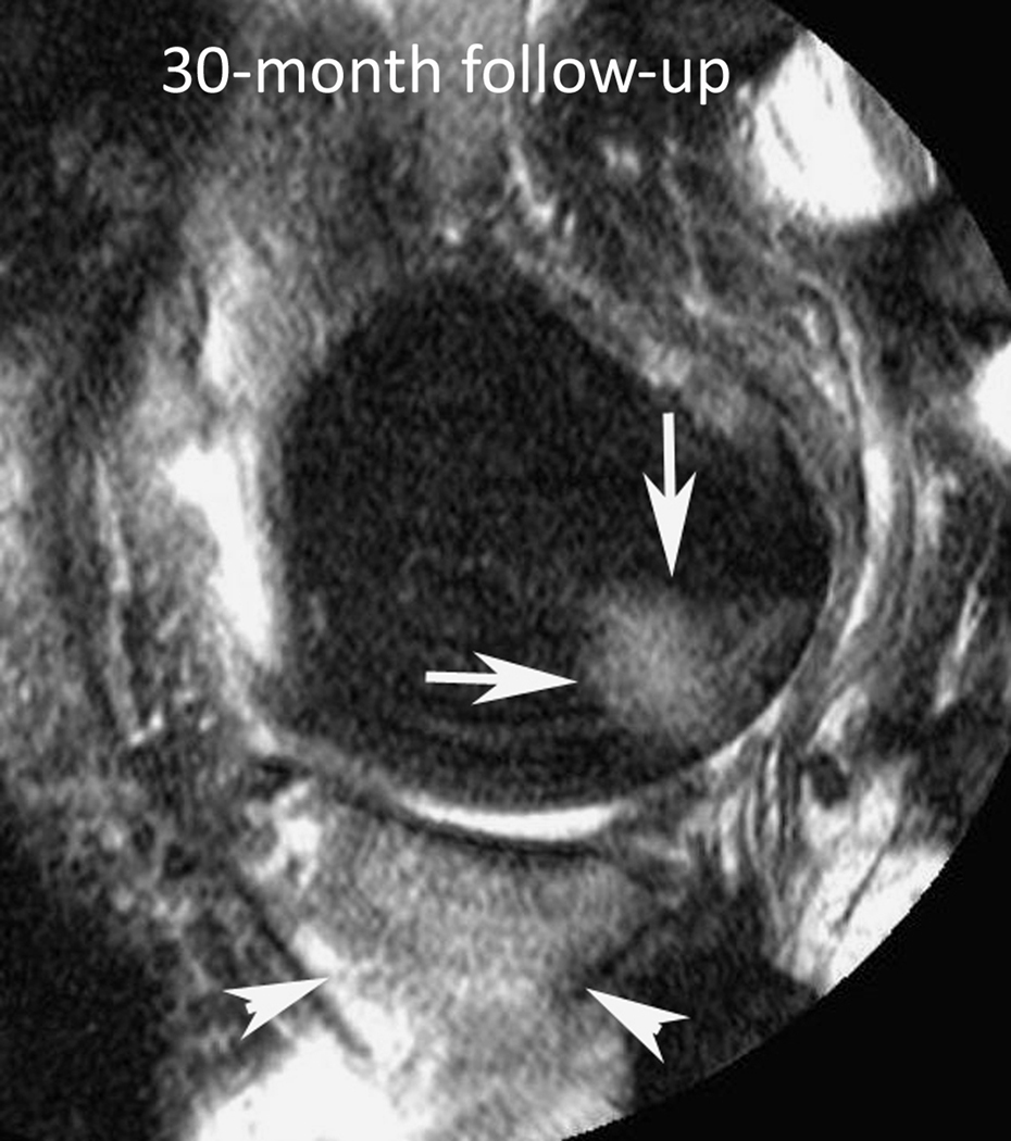
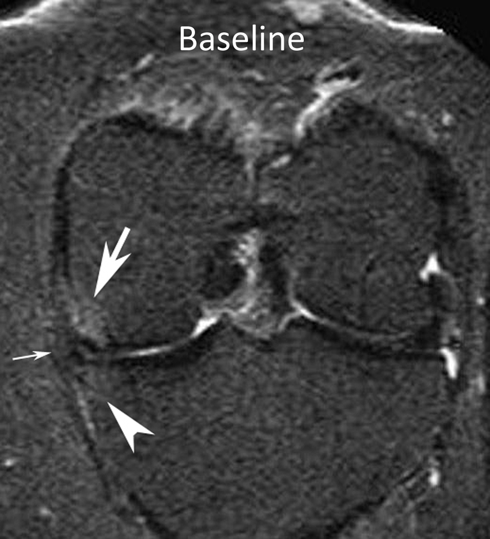
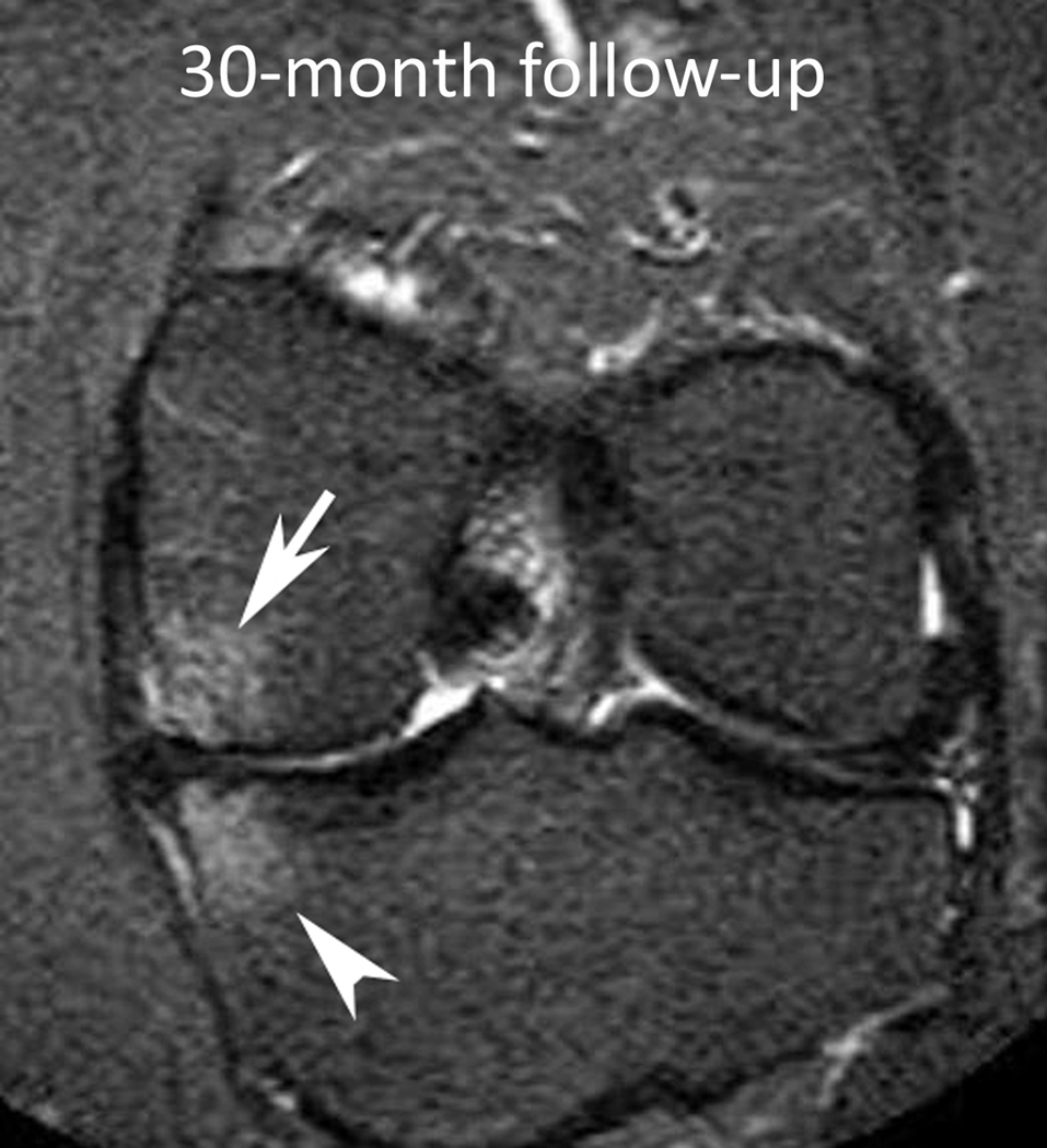
Methods: The authors studied subjects from the Multicenter Osteoarthritis Study aged 50-79 years either with knee osteoarthritis or at high risk of the disease. Baseline and 30-months magnetic resonance images of knees (n=1344) were scored for subchondral BML. Outcome was defined as an increase in BML score in either the tibial or femoral condyle in medial and lateral compartments, respectively. The authors defined meniscal pathology at baseline as the presence of either meniscal lesions or meniscal extrusion. The risk of an increase in BML score in relation to meniscal status in the same compartment was estimated using a log linear regression model adjusted for age, sex, body mass index, physical activity level and mechanical axis. In secondary analyses the investigators stratified by ipsilateral tibiofemoral cartilage status at baseline and compartments with pre-existing BML.
Results: The adjusted relative risk of incident or enlarging BML ranged from 1.8; 95% CI 1.3 to 2.3 for mild medial meniscal pathology to 5.0; 95% CI 3.2 to 7.7 for major lateral meniscal pathology (using no meniscal pathology in the same compartment as reference). Stratification by cartilage or BML status at baseline had essentially no effect on these estimates.
Conclusions: Knee compartments with meniscal pathology have a substantially increased risk of incident or enlarging subchondral BML over 30 months. Higher relative risks were seen in those with more severe and with lateral meniscal pathology.
Figures

Similar articles
-
Knee malalignment is associated with an increased risk for incident and enlarging bone marrow lesions in the more loaded compartments: the MOST study.Osteoarthritis Cartilage. 2012 Nov;20(11):1227-33. doi: 10.1016/j.joca.2012.07.020. Epub 2012 Aug 5. Osteoarthritis Cartilage. 2012. PMID: 22874524 Free PMC article.
-
Meniscal extrusion predicts increases in subchondral bone marrow lesions and bone cysts and expansion of subchondral bone in osteoarthritic knees.Rheumatology (Oxford). 2010 May;49(5):997-1004. doi: 10.1093/rheumatology/keq034. Epub 2010 Feb 24. Rheumatology (Oxford). 2010. PMID: 20181669
-
Comparison of BLOKS and WORMS scoring systems part II. Longitudinal assessment of knee MRIs for osteoarthritis and suggested approach based on their performance: data from the Osteoarthritis Initiative.Osteoarthritis Cartilage. 2010 Nov;18(11):1402-7. doi: 10.1016/j.joca.2010.06.016. Epub 2010 Sep 17. Osteoarthritis Cartilage. 2010. PMID: 20851202 Free PMC article.
-
Bone Marrow Edema: Chronic Bone Marrow Lesions of the Knee and the Association with Osteoarthritis.Bull Hosp Jt Dis (2013). 2016 Mar;74(1):24-36. Bull Hosp Jt Dis (2013). 2016. PMID: 26977546 Review.
-
The reliability of a new scoring system for knee osteoarthritis MRI and the validity of bone marrow lesion assessment: BLOKS (Boston Leeds Osteoarthritis Knee Score).Ann Rheum Dis. 2008 Feb;67(2):206-11. doi: 10.1136/ard.2006.066183. Epub 2007 May 1. Ann Rheum Dis. 2008. PMID: 17472995 Review.
Cited by
-
Predictive validity of within-grade scoring of longitudinal changes of MRI-based cartilage morphology and bone marrow lesion assessment in the tibio-femoral joint--the MOST study.Osteoarthritis Cartilage. 2012 Nov;20(11):1391-8. doi: 10.1016/j.joca.2012.07.012. Epub 2012 Jul 27. Osteoarthritis Cartilage. 2012. PMID: 22846715 Free PMC article.
-
Meniscus pathology, osteoarthritis and the treatment controversy.Nat Rev Rheumatol. 2012 May 22;8(7):412-9. doi: 10.1038/nrrheum.2012.69. Nat Rev Rheumatol. 2012. PMID: 22614907 Review.
-
Repair of Avascular Meniscus Tears with Electrospun Collagen Scaffolds Seeded with Human Cells.Tissue Eng Part A. 2016 Mar;22(5-6):436-48. doi: 10.1089/ten.TEA.2015.0284. Epub 2016 Mar 3. Tissue Eng Part A. 2016. PMID: 26842062 Free PMC article.
-
Role of Scaffolds, Subchondral, Intra-Articular Injections of Fresh Autologous Bone Marrow Concentrate Regenerative Cells in Treating Human Knee Cartilage Lesions: Different Approaches and Different Results.Int J Mol Sci. 2021 Apr 8;22(8):3844. doi: 10.3390/ijms22083844. Int J Mol Sci. 2021. PMID: 33917689 Free PMC article. Review.
-
Osteoarthritis prevention through meniscal regeneration induced by intra-articular injection of meniscus stem cells.Stem Cells Dev. 2013 Jul 15;22(14):2071-82. doi: 10.1089/scd.2012.0563. Epub 2013 Apr 1. Stem Cells Dev. 2013. PMID: 23461527 Free PMC article.
References
-
- Fukubayashi T, Kurosawa H. The contact area and pressure distribution pattern of the knee. A study of normal and osteoarthrotic knee joints. Acta Orthop Scand. 1980;51:871–879. - PubMed
-
- Seedhom BB, Hargreaves DJ. Transmission of the load in the knee joint with special reference to the role of the meniscus. Part I+II. Eng Med. 1979;4:207–228.
-
- Walker PS, Erkman MJ. The role of the menisci in force transmission across the knee. Clin Orthop Relat Res. 1975:184–192. - PubMed
-
- Englund M, Lohmander LS. Risk factors for symptomatic knee osteoarthritis fifteen to twenty-two years after meniscectomy. Arthritis Rheum. 2004;50:2811–2819. - PubMed
Publication types
MeSH terms
Grants and funding
LinkOut - more resources
Full Text Sources
Other Literature Sources
Medical
