. 2010 Jul;30(13):3371-83.
doi: 10.1128/MCB.01644-09. Epub 2010 Apr 26.

Role of ATM and the damage response mediator proteins 53BP1 and MDC1 in the maintenance of G(2)/M checkpoint arrest

Affiliations

Role of ATM and the damage response mediator proteins 53BP1 and MDC1 in the maintenance of G(2)/M checkpoint arrest

Atsushi Shibata et al. Mol Cell Biol. 2010 Jul.

Abstract

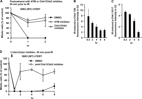
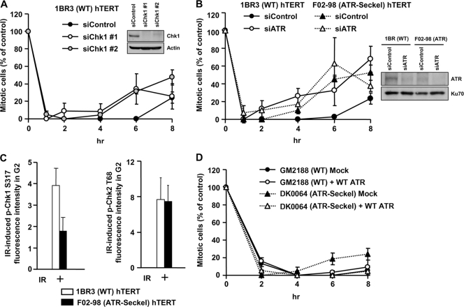
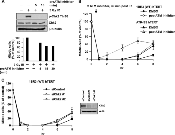
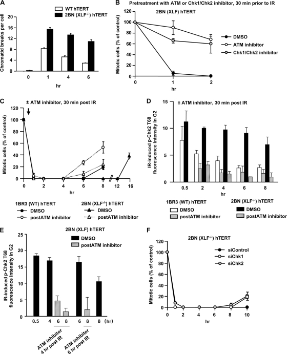
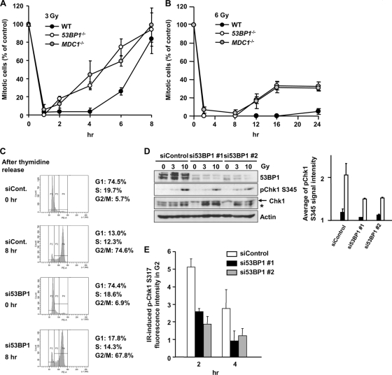
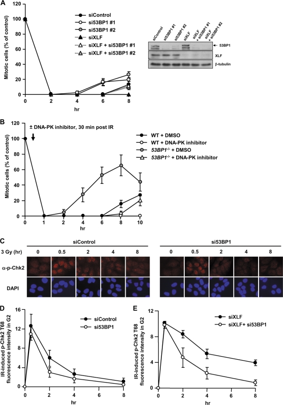
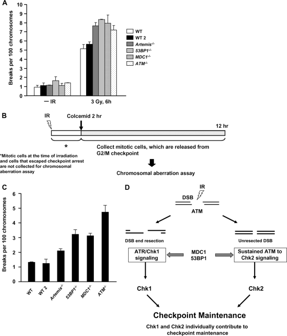
ATM-dependent initiation of the radiation-induced G(2)/M checkpoint arrest is well established. Recent results have shown that the majority of DNA double-strand breaks (DSBs) in G(2) phase are repaired by DNA nonhomologous end joining (NHEJ), while approximately 15% of DSBs are slowly repaired by homologous recombination. Here, we evaluate how the G(2)/M checkpoint is maintained in irradiated G(2) cells, in light of our current understanding of G(2) phase DSB repair. We show that ATM-dependent resection at a subset of DSBs leads to ATR-dependent Chk1 activation. ATR-Seckel syndrome cells, which fail to efficiently activate Chk1, and small interfering RNA (siRNA) Chk1-treated cells show premature mitotic entry. Thus, Chk1 significantly contributes to maintaining checkpoint arrest. Second, sustained ATM signaling to Chk2 contributes, particularly when NHEJ is impaired by XLF deficiency. We also show that cells lacking the mediator proteins 53BP1 and MDC1 initially arrest following radiation doses greater than 3 Gy but are subsequently released prematurely. Thus, 53BP1(-/-) and MDC1(-/-) cells manifest a checkpoint defect at high doses. This failure to maintain arrest is due to diminished Chk1 activation and a decreased ability to sustain ATM-Chk2 signaling. The combined repair and checkpoint defects conferred by 53BP1 and MDC1 deficiency act synergistically to enhance chromosome breakage.


Figures

FIG. 1.
FIG. 1.
Chk1/Chk2 regulates the initiation and maintenance of checkpoint arrest. (A) Checkpoint initiation requires ATM and Chk1/Chk2. Mitotic entry was examined in 1BR3 hTERT cells following 3 Gy IR in untreated cells or cells treated with the ATM inhibitor or Chk1/Chk2 inhibitor 30 min prior to IR. Cells, >400, were scored for mitotic index. Results represent the mitotic index relative to untreated cells. Maximal levels of p-Chk1 and p-Chk2 are reached within 30 min after 3 Gy. 1BR hTERT and A549 cells were exposed to 3 Gy IR, and p-Chk2 (B) and p-Chk1 (C) levels assessed by IF at various times post-IR. (D) Checkpoint maintenance requires Chk1/Chk2. The time of mitotic entry following exposure to 3 Gy IR in cells either untreated or treated with the Chk1/Chk2 inhibitor 30 min post-IR was examined. Error bars represent the standard error of the mean (SEM) from 3 experiments.
FIG. 2.
FIG. 2.
ATR-Chk1 signaling contributes to checkpoint maintenance. (A) Mitotic entry in 1BR3 hTERT cells following treatment with control and Chk1 siRNA and 3 Gy IR was examined. Chk1 knockdown did not significantly compromise G2 proportion based on fluorescence-activated cell sorting (FACS) analysis (data not shown). (B) Mitotic entry in 1BR3 hTERT and ATR-SS hTERT cells following treatment with control or ATR siRNA and 3 Gy IR. The right panel shows ATR expression levels. (C) ATR-SS hTERT cells show impaired Chk1 phosphorylation but normal Chk2 phosphorylation. The levels of p-Chk1 Ser317 and p-Chk2 Thr68 were quantified by IF in ATR-SS hTERT G2 cells at 30 min after IR exposure. (D) Mitotic entry in control and ATR-SS LBL cell lines following mock and ATR cDNA transfection. To verify the impact of Chk1 siRNA on Chk1 activity, we examined hydroxyurea (HU)-induced 53BP1 focus formation, which is a characterized Chk1-dependent response (26). 53BP1 foci failed to form after HU treatment in ATR-SS hTERT cells and following Chk1 siRNA, consistent with previous findings (data not shown) (30). Error bars represent the SEM from 3 experiments.
FIG. 3.
FIG. 3.
Sustained ATM signaling also contributes to checkpoint maintenance. (A) ATM inhibitor addition impairs checkpoint arrest and signaling within 5 min. 1BR3 hTERT cells were untreated or treated with ATM inhibitor 5, 15, or 30 min prior to IR. Cells were examined by immunoblotting using anti-Thr68-p-Chk2 at 30 min (top) and for mitotic entry at 1 h post-IR (bottom). Chk2 and β-tubulin antibodies were loading controls. (B) ATM inhibitor addition 30 min post-IR diminishes the duration of checkpoint arrest after 3 Gy IR in 1BR3 hTERT and in ATR-SS hTERT cells. (C) siRNA-Chk2 diminishes the duration of checkpoint arrest after 3 Gy IR. Error bars represent the SEM of 3 experiments.
FIG. 4.
FIG. 4.
Sustained ATM signaling contributes to prolonged checkpoint arrest in NHEJ-defective cells. (A) Cells were exposed to 1 Gy IR, and APH was added. A total of 50 ng/ml calyculin A was added 30 min before fixation, and PCCs were scored. (B) ATM and Chk1/Chk2 are required for checkpoint initiation in 2BN (XLF−/−) hTERT cells. Mitotic entry was examined following 3 Gy IR in 2BN (XLF−/−) hTERT cells either untreated or treated with the ATM or Chk1/Chk2 inhibitor 30 min prior to IR. (C) Prolonged checkpoint arrest in 2BN (XLF−/−) hTERT cells requires sustained ATM signaling. Mitotic entry after 3 Gy IR was examined in 1BR3 and 2BN (XLF−/−) hTERT cells either with or without ATM inhibitor 30 min post-IR. (D) 1BR3 and 2BN (XLF−/−) hTERT cells were exposed to 3 Gy IR with or without ATM inhibitor addition 30 min post-IR. p-Chk2 levels were quantified in CENP-F+ (G2) cells by IF using anti-Thr68-p-Chk2 antibodies. The background signal in unirradiated cells was subtracted. At <1 h post-IR, 2BN (XLF−/−) hTERT cells harbor elevated DSBs compared to 1BR3 hTERT cells (as shown in panel A), consistent with their elevated p-Chk2 signal. By 8 h the p-Chk2 level decreased by >50% in 1BR3 hTERT cells compared with 20% in 2BN (XLF−/−) hTERT cells. (E) 2BN (XLF−/−) hTERT cells were exposed to 3 Gy IR with or without ATM inhibitor addition 4 or 6 h post-IR. p-Chk2 levels were quantified in CENP-F+ (G2) cells by IF using anti-Thr68-p-Chk2 antibodies. (F) The maintenance of checkpoint arrest in 2BN (XLF−/−) hTERT cells requires Chk1 and Chk2. Mitotic entry was examined in 2BN (XLF−/−) hTERT cells treated with control, Chk1, or Chk2 siRNA. Error bars represent the SEM of 3 experiments.
FIG. 5.
FIG. 5.
MDC1−/ and 53BP1−/− MEFs show impaired G2/M checkpoint maintenance and reduced Chk1 phosphorylation. (A and B) Mitotic entry in WT, MDC1−/−, and 53BP1−/− MEFs after 3 Gy and 6 Gy IR was evaluated. APH was added after IR. (C) Cell cycle profile in control and 53BP1 siRNA cells after double thymidine block. (D) p-Chk1 is reduced in synchronized G2 53BP1 siRNA cells post-IR. Cells were synchronized in G1/S by double thymidine block and irradiated 8 h postrelease. Cells were examined by immunoblotting using anti-pChk1 Ser345 antibody. The asterisk represents a nonspecific band. Quantification of p-Chk1 from 2 experiments is shown. (E) 53BP1 siRNA cells show impaired Chk1 activation. The p-Ser317 Chk1 signal was quantified by IF in CENP-F+ (G2) A549 cells treated 3 Gy IR and 53BP1 or control siRNA. The signal in undamaged nuclei is subtracted. Error bars represent the SEM of 3 experiments.
FIG. 6.
FIG. 6.
53BP1-defective cells show impaired sustained ATM-Chk2 signaling. (A) A549 cells treated with 53BP1 siRNA, XLF siRNA, or both were exposed to 3 Gy IR, and the mitotic entry was evaluated. 53BP1 and XLF siRNA causes prolonged arrest compared to 53BP1 siRNA alone. The knockdown efficiency is shown in right panel. (B) WT or 53BP1−/− MEFs were treated with or without DNA-PK inhibitor addition 30 min after 3 Gy IR, and mitotic entry was examined. (C) Typical picture of p-Chk2 Thr68 in 53BP1 siRNA A549 cells after 3 Gy IR. α, anti. (D) Quantification of p-Chk2 levels in 53BP1 siRNA-treated A549 cells after 3 Gy IR. p-Chk2 levels were assessed by IF quantification in CENP-F+ (G2) cells. (E) Quantification of the p-Chk2 in G2 cells following XLF or 53BP1/XLF siRNA after 3 Gy IR.
FIG. 7.
FIG. 7.
The combined DSB repair and signaling defects in MDC1−/ and 53BP1−/− MEFs contribute to chromosome breakage. (A) Cells were exposed to 3 Gy IR. A total of 50 ng/ml calyculin A was added 20 min before fixation. PCCs are scored per 100 chromosomes. (B) Diagram showing the procedure used. Colcemid was added from 2 to 12 h after 3 Gy IR. Mitotic cells were not collected during the first 2 h post-IR to prevent analysis of cells that “escape” checkpoint arrest. Chromatid breaks were scored. (C) Mitotic chromosomal break analysis of 2 WT, MDC1−/−, 53BP1−/−, Artemis−/−, and ATM−/− MEFs. Chromatid break numbers were normalized to 100 chromosomes to account for differences in chromosome numbers for different lines. (D) Model showing the mechanisms contributing to checkpoint maintenance and the contribution of 53BP1 and MDC1.

Similar articles

Cited by

References

    1. Ahnesorg, P., P. Smith, and S. P. Jackson. 2006. XLF interacts with the XRCC4-DNA ligase IV complex to promote DNA nonhomologous end-joining. Cell 124:301-313. - PubMed
    1. Beamish, H., and M. F. Lavin. 1994. Radiosensitivity in ataxia-telangiectasia: anomalies in radiation-induced cell cycle delay. Int. J. Radiat. Biol. 65:175-184. - PubMed
    1. Beucher, A., J. Birraux, L. Tchouandong, O. Barton, A. Shibata, S. Conrad, A. A. Goodarzi, A. Krempler, P. A. Jeggo, and M. Lobrich. 2009. ATM and Artemis promote homologous recombination of radiation-induced DNA double-strand breaks in G2. EMBO J. 28:3413-3427. - PMC - PubMed
    1. Buck, D., L. Malivert, R. de Chasseval, A. Barraud, M. C. Fondaneche, O. Sanal, A. Plebani, J. L. Stephan, M. Hufnagel, F. le Deist, A. Fischer, A. Durandy, J. P. de Villartay, and P. Revy. 2006. Cernunnos, a novel nonhomologous end-joining factor, is mutated in human immunodeficiency with microcephaly. Cell 124:287-299. - PubMed
    1. Dai, Y., B. Kysela, L. A. Hanakahi, K. Manolis, E. Riballo, M. Stumm, T. O. Harville, S. C. West, M. A. Oettinger, and P. A. Jeggo. 2003. Non-homologous end joining and V(D)J recombination require an additional factor. Proc. Natl. Acad. Sci. U. S. A. 100:2462-2467. - PMC - PubMed

Publication types

MeSH terms

Substances

LinkOut - more resources