. 2010 Jul;30(13):3176-86.
doi: 10.1128/MCB.00214-10. Epub 2010 Apr 26.

Myogenic microRNA expression requires ATP-dependent chromatin remodeling enzyme function

Affiliations

Myogenic microRNA expression requires ATP-dependent chromatin remodeling enzyme function

Chandrashekara Mallappa et al. Mol Cell Biol. 2010 Jul.

Abstract

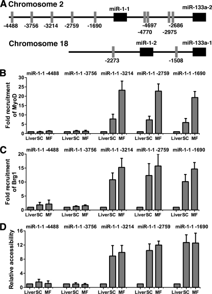
Knockdown of the Brg1 ATPase subunit of SWI/SNF chromatin remodeling enzymes in developing zebrafish caused stunted tail formation and altered sarcomeric actin organization, which phenocopies the loss of the microRNA processing enzyme Dicer, or the knockdown of myogenic microRNAs. Furthermore, myogenic microRNA expression and differentiation was blocked in Brg1 conditional myoblasts differentiated ex vivo. The binding of Brg1 upstream of myogenic microRNA sequences correlated with MyoD binding and accessible chromatin structure in satellite cells and myofibers, and it was required for chromatin accessibility and microRNA expression in a tissue culture model for myogenesis. The results implicate ATP-dependent chromatin remodelers in myogenic microRNA gene regulation.


Figures

FIG. 1.
FIG. 1.
Brg1-MO injection alters tail skeletal muscle organization. Zebrafish embryos were injected with control MO (A, C, E, and G) or Brg1-MO (B, D, F, and H), and animals were imaged at 28 hpf. (A and B) Normal and stunted tail development. (C) Normal somite structure was observed following control MO injection. (D) Altered somite structure was observed following Brg1-MO injection. Scale bar, 100 μm. (E and F) MyHC staining in the tail following control or Brg1-MO injection. Scale bar, 50 μm. (G and H) Tail skeletal muscle was immunostained for α-actin following control or Brg1-MO injection. Note the loss of I band staining in the sarcomeres in the Brg1-MO-injected tissue. Scale bar, 10 μm.
FIG. 2.
FIG. 2.
Brg1-MO-injected zebrafish with altered tail development have compromised α-actin and skeletal muscle microRNA expression. (A) qPCR analyses of α-actin, Dicer, and miRNA-29a primary transcript levels in control- or Brg1-MO-injected animals. (B) qPCR to detect the indicated miRNA primary transcript levels in control MO-injected embryos or in Brg1-MO-injected animals that exhibited a tail phenotype. The expression of each gene in the control MO-injected animals was normalized to 1; results are the averages from three independent experiments performed on different days ± standard deviations. For each independent experiment, eight or nine embryos were pooled for RNA preparation.
FIG. 3.
FIG. 3.
Brg1 conditional myoblasts treated with Ad-Cre do not differentiate. (A) Myoblasts isolated from newborn Brg1 conditional mice were cultured, mock infected, or infected with 0.5 μl (1,125 PFU/cell) Ad-Cre, and then they were induced to differentiate by the addition of low-serum media. Four days postdifferentiation, cells were stained for MyHC and with 4′,6′-diamidino-2-phenylindole (DAPI) and compared to bright-field images. (B) The same experiment as that described for panel A was repeated with myoblasts isolated from wild-type mice (top and middle) or with Brg1 conditional myoblasts infected with 5 μl (3,750 PFU/cell) of Ad-lacZ (bottom). Scale bar, 50 μm.
FIG. 4.
FIG. 4.
Brg1 conditional myoblasts treated with Ad-Cre are impaired in myogenic microRNA expression. (A) Excision of the Brg1 conditional allele as a function of an increasing dose of Ad-Cre (0, 0.1, 0.5, 1, and 2 μl; 1 μl = 2,250 PFU/cell). (B) Western blot demonstrating Brg1 protein levels in Brg1 conditional myoblasts treated with 0, 0.5, or 1 μl of Ad-Cre. PI3 kinase (PI3K) levels were monitored as a control. (C) Relative miRNA primary transcript levels present in Brg1 conditional myoblasts infected with increasing amounts of Ad-Cre (0, 0.1, 0.5, 1, and 2 μl) and assayed at day 4 postdifferentiation. (D) Relative expression of miR-1 and miR-133a primary transcripts in wild-type myoblasts infected with 0, 1, or 2 μl of Ad-Cre and assayed at day 0 or 4 postdifferentiation. (E) Relative expression of miR-1 and miR-133a primary transcripts from Brg1 conditional myoblasts infected with Ad-LacZ (0, 5, or 10 μl; 5 μl = 3,750 PFU/cell) and assayed at day 0 or 4 postdifferentiation. MicroRNA primary transcript levels presented in panels C to E were quantified in three independent experiments and are presented as averages ± standard deviations. The expression in the absence of Ad-Cre or Ad-lacZ was normalized to 1.
FIG. 5.
FIG. 5.
Myogenic microRNAs are highly induced in primary skeletal muscle tissues. (A) Relative expression levels of miRNA primary transcripts miR-1-1, miR-1-2, miR-133a-1, and miR-133a-2 in liver (L), satellite cells (SC), or myofibers (MF). Transcript levels in liver tissue were normalized to 1. (B and C) Northern blots of mature miR-1 and miR-133a levels in these tissues. U6 snRNA was measured as a loading control.
FIG. 6.
FIG. 6.
Brg1 binding correlates with chromatin remodeling near all but the most distal of the E boxes upstream of the miR-1-1 stem-loop sequence in primary tissue. (A) Schematic maps of the miR-1 and miR-133 loci. The positions of consensus E boxes upstream of each miRNA stem-loop sequence are indicated. (B and C) ChIP experiments demonstrating the binding of MyoD (B) and Brg1 (C) near all but the most distal E boxes from the miR-1-1 stem-loop sequence in satellite cells (SC) and myofibers (MF) but not liver tissue. Levels of MyoD or Brg1 binding at each sequence in the liver sample were normalized to 1. (D) REAA from the tissue samples used for panels B and C indicating increased nuclease accessibility at PvuII sites (which exactly correspond to E boxes) at all but the most distal E boxes from the miR-1-1 stem-loop sequence. Enzyme cleavage at each sequence in the liver sample was normalized to 1. Data in panels B to D are the averages from three independent tissue isolations ± standard deviations.
FIG. 7.
FIG. 7.
MyoD binding, Brg1 binding, and chromatin accessibility correlate at E box regions of miR-1-2, miR-133a-1, and miR-133a-2 in primary tissue. MyoD (A) and Brg1 (B) recruitment at the indicated E box-containing sequences upstream of the miR-1-2, miR-133a-1, and miR-133a-2 coding sequences in liver (L), satellite cells (SC), or myofibers (MF). Levels of MyoD or Brg1 binding at each sequence in the liver sample were normalized to 1. Background binding observed with IgG pulldown in each tissue is shown as a control. (C) REAA at the indicated E boxes upstream of the miR-1-2, miR-133a-1, and miR-133a-2 coding sequences in liver (L), satellite cells (SC), or myofibers (MF). Enzyme cleavage at each sequence in the liver sample was normalized to 1. Accessibility at the E box in the Eef1 α1 coding region was examined as a control. Data are the averages from three independent tissue isolations ± standard deviations.
FIG. 8.
FIG. 8.
Expression of miRNAs miR-1 and miR-133a is compromised in differentiated cells expressing DN Brg1. B22 cells express dominant-negative Brg1 in the absence but not the presence of tetracycline (Tet). B22 cells cultured in the presence or absence of Tet were infected with the pBABE retroviral vector (V) or pBABE-MyoD (D), and samples were collected at the onset of differentiation, designated time zero, and subsequently differentiated by the addition of a low-serum medium for 28 h. (A and B) Northern blots for miR-1 and miR-133a miRNAs in differentiated B22 cells. (C) Expression of the widely expressed miR-29a miRNA was monitored by Northern blotting as a control. U6 snRNA levels were measured as a loading control for each blot.
FIG. 9.
FIG. 9.
Functional Brg1 is required for chromatin remodeling at and gene expression from the miR-1-1 locus in cultured cells. (A) B22 cells cultured without tetracycline (Tet) express DN Brg1. Cells were infected with empty retroviral pBABE vector (V) or with a MyoD-encoding retrovirus (D) and were collected at the onset of differentiation, which was designated 0 h, or at 28 h postdifferentiation. Cells expressing DN Brg1 showed significantly reduced levels of miR-1-1, miR-1-2, miR-133a-1, and miR-133a-2 primary transcripts compared to those of cells grown with Tet. (B and C) ChIP experiments examining the presence of MyoD or Brg1 near E boxes upstream of the miR-1-1 gene in the samples described for panel A. (D) REAA experiments examining chromatin accessibility in these samples at the indicated E boxes. All experiments are the averages of three independent experiments ± standard deviations, and expression, binding, or enzyme cleavage at each sequence in the vector-differentiated cells plus tetracycline at time zero was normalized to 1.
FIG. 10.
FIG. 10.
Expression of DN Brg1 affects Brg1 binding and chromatin accessibility at upstream E boxes of miR-1-2, miR-133a-1, and miR-133a-2 stem-loop sequences. MyoD (A) and Brg1 (B) ChIPs at E box-containing sequences upstream of the miR-1-2, miR-133a-1, and miR-133a-2 gene sequences in B22 cells treated as described for Fig. 9. (C) REAA at the indicated E boxes upstream of the miR-1-2, miR-133a-1, and miR-133a-2 genes. Binding or enzyme cleavage at each sequence in the vector-differentiated cells plus tetracycline at time zero was normalized to 1. Data represent the averages from three independent experiments ± standard deviations.

Similar articles

Cited by

References

    1. Andl, T., E. P. Murchison, F. Liu, Y. Zhang, M. Yunta-Gonzalez, J. W. Tobias, C. D. Andl, J. T. Seykora, G. J. Hannon, and S. E. Millar. 2006. The miRNA-processing enzyme dicer is essential for the morphogenesis and maintenance of hair follicles. Curr. Biol. 16:1041-1049. - PMC - PubMed
    1. Bartel, D. P. 2004. MicroRNAs: genomics, biogenesis, mechanism, and function. Cell 116:281-297. - PubMed
    1. Berkes, C. A., D. A. Bergstrom, B. H. Penn, K. J. Seaver, P. S. Knoepfler, and S. J. Tapscott. 2004. Pbx marks genes for activation by MyoD indicating a role for a homeodomain protein in establishing myogenic potential. Mol. Cell 14:465-477. - PubMed
    1. Bernstein, E., S. Y. Kim, M. A. Carmell, E. P. Murchison, H. Alcorn, M. Z. Li, A. A. Mills, S. J. Elledge, K. V. Anderson, and G. J. Hannon. 2003. Dicer is essential for mouse development. Nat. Genet. 35:215-217. - PubMed
    1. Bultman, S., T. Gebuhr, D. Yee, C. La Mantia, J. Nicholson, A. Gilliam, F. Randazzo, D. Metzger, P. Chambon, G. Crabtree, and T. Magnuson. 2000. A Brg1 null mutation in the mouse reveals functional differences among mammalian SWI/SNF complexes. Mol. Cell 6:1287-1295. - PubMed

Publication types

MeSH terms

LinkOut - more resources