

doi: 10.1104/pp.110.156844.
Epub 2010 Apr 26.
The XTH gene family: an update on enzyme structure, function, and phylogeny in xyloglucan remodeling
Affiliations
- PMID: 20421457
- PMCID: PMC2879796
- DOI: 10.1104/pp.110.156844
Item in Clipboard
The XTH gene family: an update on enzyme structure, function, and phylogeny in xyloglucan remodeling
Plant Physiol.
2010 Jun.
No abstract available
Figures
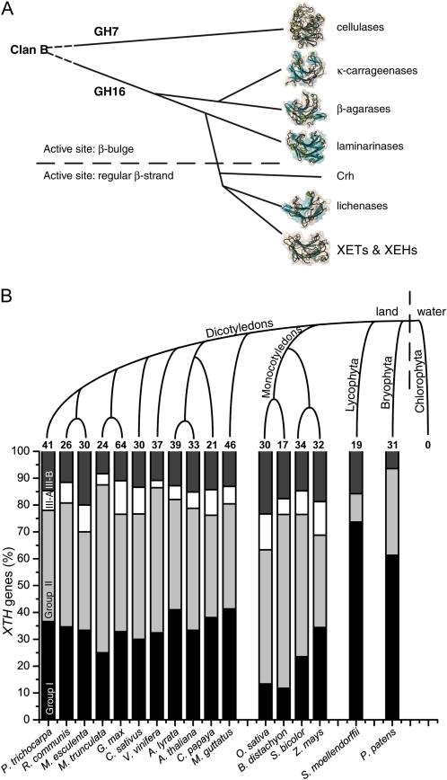
The evolution of GH16 and grouping of XTH genes in publicly available plant genomes. A, The proposed evolution of clan B containing GH7 and GH16 (updated from Michel et al. [2001]). B, A simplified tree showing the genome representatives of the Plantae (top) and a diagram showing the distribution of XTH genes into groups (bottom), with the number of genes from each organism at the top of each column (only XTH genes predicted to encode functional XTH gene products [i.e. containing a complete active-site motif] are included). The groups are colored as follows: group 1, black; group II, light gray; group III-A, white; group III-B, dark gray. The Chlorophyta are represented by the genomes of Chlamydomonas reinhardtii (http://phytozome.org/ ), Micromonas pusilla (Worden et al., 2009), Ostreococcus tauri (Derelle et al., 2006), and Ostreococcus lucimarinus (Palenik et al., 2007). All other genome data are available via http://www.phytozome.org/ (accessed March 2010).
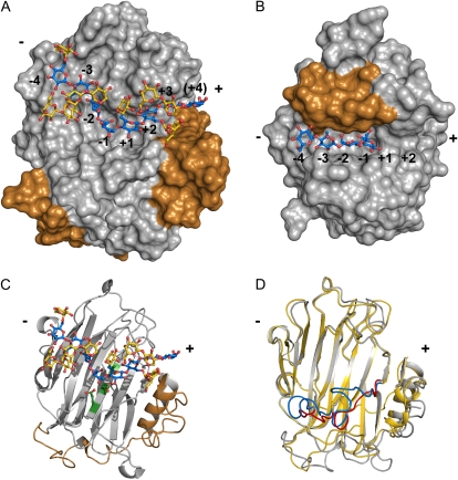
Structures of XTH gene products and a closely related GH16 β-1,3;1,4-glucanase. A, Surface representation of PttXET16-34 (PDB ID 1un1, 1umz) in silver (C-terminal extension in copper) with XLLGXLLG octadecasaccharide bound (glucosyl backbone in blue and xylosyl and galactosyl units in gold; based on data from Mark et al. [2009]). B, Surface representation of a β-1,3;1,4-glucanase in silver, with the loop narrowing the negative subsites in copper (PDB ID 1u0a). C, Cartoon of PttXET16-34 showing the structure of the C-terminal extension (copper) and the catalytic amino acids (green) with a bound XLLGXLLG octadecasaccharide. D, Overlay of a XET, PttXET16-34 (silver with red loops), and an XEH, TmNXG1 (gold with blue loops; PDB ID 2uwa, 2vh9).
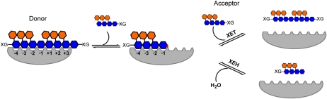
A schematic representation of the mechanism used by XETs and XEHs. Left, xyloglucan binds to XETs and XEHs in both negative and positive subsites (glucosyl units of xyloglucan in blue and xylosyl units in orange; subsite nomenclature of Davies et al. [1997]). After binding, the substrate is cleaved, resulting in a covalent glycosyl-enzyme intermediate (bond indicated in red). Right, in the last step, the glycosyl-enzyme is broken down by an incoming acceptor, either water (XEH activity) or the nonreducing end of a xyloglucan molecule (XET activity).
Similar articles
-
Unraveling the functions of glycosyltransferase family 47 in plants.Trends Plant Sci. 2003 Dec;8(12):565-8. doi: 10.1016/j.tplants.2003.10.003. Trends Plant Sci. 2003. PMID: 14659703
-
The XTH family of enzymes involved in xyloglucan endotransglucosylation and endohydrolysis: current perspectives and a new unifying nomenclature.Plant Cell Physiol. 2002 Dec;43(12):1421-35. doi: 10.1093/pcp/pcf171. Plant Cell Physiol. 2002. PMID: 12514239 Review.
-
Xyloglucan endotransglycosylase, a new wall-loosening enzyme activity from plants.Biochem J. 1992 Mar 15;282 ( Pt 3)(Pt 3):821-8. doi: 10.1042/bj2820821. Biochem J. 1992. PMID: 1554366 Free PMC article.
-
Another building block in the plant cell wall: Barley xyloglucan xyloglucosyl transferases link covalently xyloglucan and anionic oligosaccharides derived from pectin.Plant J. 2020 Nov;104(3):752-767. doi: 10.1111/tpj.14964. Epub 2020 Sep 19. Plant J. 2020. PMID: 32799357
-
Broad Specific Xyloglucan:Xyloglucosyl Transferases Are Formidable Players in the Re-Modelling of Plant Cell Wall Structures.Int J Mol Sci. 2022 Jan 31;23(3):1656. doi: 10.3390/ijms23031656. Int J Mol Sci. 2022. PMID: 35163576 Free PMC article. Review.
Cited by
-
Nematode feeding sites: unique organs in plant roots.Planta. 2013 Nov;238(5):807-18. doi: 10.1007/s00425-013-1923-z. Epub 2013 Jul 4. Planta. 2013. PMID: 23824525 Review.
-
Genome sequencing of Elaeocarpus spp. stem blight pathogen Pseudocryphonectria elaeocarpicola reveals potential adaptations to colonize woody bark.BMC Genomics. 2024 Jul 24;25(1):714. doi: 10.1186/s12864-024-10615-5. BMC Genomics. 2024. PMID: 39048950 Free PMC article.
-
Analysis of xyloglucan endotransglycosylase/hydrolase (XTH) genes and diverse roles of isoenzymes during persimmon fruit development and postharvest softening.PLoS One. 2015 Apr 7;10(4):e0123668. doi: 10.1371/journal.pone.0123668. eCollection 2015. PLoS One. 2015. PMID: 25849978 Free PMC article.
-
Identifying plant genes shaping microbiota composition in the barley rhizosphere.Nat Commun. 2022 Jun 16;13(1):3443. doi: 10.1038/s41467-022-31022-y. Nat Commun. 2022. PMID: 35710760 Free PMC article.
-
Effect of wet storage conditions on potato tuber transcriptome, phytohormones and growth.BMC Plant Biol. 2019 Jun 17;19(1):262. doi: 10.1186/s12870-019-1875-y. BMC Plant Biol. 2019. PMID: 31208336 Free PMC article.
References
-
- Albersheim P. (1976) The primary cell wall. Bonner J, Varner JE, , Plant Biochemistry. Academic Press, New York, pp 225–274
-
- Atkinson RG, Johnston SL, Yauk YK, Sharma NN, Schroder R. (2009) Analysis of xyloglucan endotransglucosylase/hydrolase (XTH) gene families in kiwifruit and apple. Postharvest Biol Technol 51: 149–157
-
- Becnel J, Natarajan M, Kipp A, Braam J. (2006) Developmental expression patterns of Arabidopsis XTH genes reported by transgenes and Genevestigator. Plant Mol Biol 61: 451–467 - PubMed
Publication types
MeSH terms
Substances
LinkOut - more resources
Full Text Sources
