. 2010 May 11;107(19):8639-43.
doi: 10.1073/pnas.0911647107. Epub 2010 Apr 26.

Unique multipotent cells in adult human mesenchymal cell populations

Affiliations

Unique multipotent cells in adult human mesenchymal cell populations

Yasumasa Kuroda et al. Proc Natl Acad Sci U S A. .

Abstract

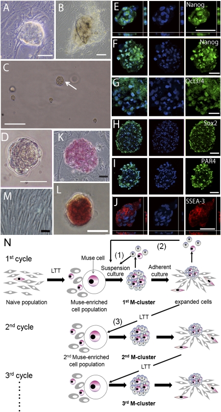
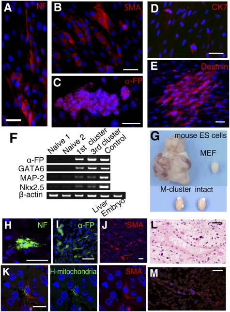
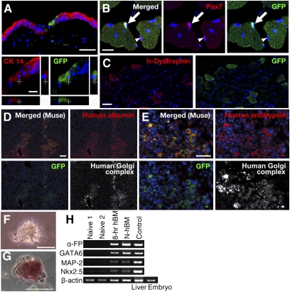
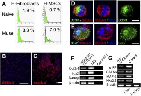
We found adult human stem cells that can generate, from a single cell, cells with the characteristics of the three germ layers. The cells are stress-tolerant and can be isolated from cultured skin fibroblasts or bone marrow stromal cells, or directly from bone marrow aspirates. These cells can self-renew; form characteristic cell clusters in suspension culture that express a set of genes associated with pluripotency; and can differentiate into endodermal, ectodermal, and mesodermal cells both in vitro and in vivo. When transplanted into immunodeficient mice by local or i.v. injection, the cells integrated into damaged skin, muscle, or liver and differentiated into cytokeratin 14-, dystrophin-, or albumin-positive cells in the respective tissues. Furthermore, they can be efficiently isolated as SSEA-3(+) cells. Unlike authentic ES cells, their proliferation activity is not very high and they do not form teratomas in immunodeficient mouse testes. Thus, nontumorigenic stem cells with the ability to generate the multiple cell types of the three germ layers can be obtained through easily accessible adult human mesenchymal cells without introducing exogenous genes. These unique cells will be beneficial for cell-based therapy and biomedical research.


Conflict of interest statement

The authors declare no conflict of interest.

Figures

Fig. 1.
Fig. 1.
Characterization of M-clusters. (A and B) Characteristic cell clusters that occur spontaneously in adherent cultures of naive H-MSCs. (C and D) MC culture of H-fibroblasts on day 7 showing an M-cluster (C, arrow). Immunocytochemical localization of Nanog (E and F), Oct3/4 (G), Sox2 (H), PAR4 (I), and SSEA-3 (J) in M-clusters formed by H-fibroblasts (E, I, and J) and H-MSCs (F, G, and H). ALP(+) human ES cells (K), M-cluster (H-fibroblast) (L), and naive H-fibroblasts (M). (N) Schematic diagram of the self-renewal of Muse cells. (Scale bars: AC, 100 μm; DM, 50 μm.)
Fig. 2.
Fig. 2.
Differentiation of Muse cells in vitro and in testes. Immunocytochemistry of neurofilament-M (NF) (A), α-SMA (B), α-fetoprotein (α-FP) (C), cytokeratin 7 (CK7) (D), and desmin (E) in cells derived from a single M-cluster (H-fibroblasts). (F) RT-PCR analysis of naive cells and first- and third-generation M-clusters (first and third clusters) derived from H-fibroblasts. Positive controls were human fetus liver (Liver) for α-FP and whole human embryo (Embryo) for GATA6, MAP-2, and Nkx2.5. (GM) Testes of immunodeficient mice injected with cells. (G) Uninjected testes (intact) and testes injected with mouse ES cells (8 weeks), mouse embryonic fibroblast (MEF) cells (8 weeks), and M-clusters (6 months). Immunohistochemistry of NF (H), α-FP (I), and SMA (J) in testes injected with MEC populations and M-clusters. (K) Double-labeling of human mitochondria (green) and SMA (red). The tube-like structure (L) was positive for human mitochondria in the adjacent section (M; red). (Scale bars: AE and HL, 50 μm; M, 20 μm.)
Fig. 3.
Fig. 3.
Transplantation of Muse cells and M-cluster formation from bone marrow. (AE) Differentiation of GFP-labeled MEC population (H-fibroblasts) in damaged tissues of immunodeficient mice. (A) Cells locally injected into the edge of the excised region. Transplanted GFP(+) cells expressed cytokeratin 14 (red) in the regenerating epidermis (2 weeks). (B) Two weeks after i.v. injection, GFP(+) cells with central nuclei were seen in cardiotoxin-injected cutaneous muscle. Transplanted GFP(+) cells (arrow) and host cells [GFP(−), arrowhead] that expressed Pax7 were seen. (C) After 4 weeks, the GFP(+) muscle fibers expressed human dystrophin (h-Dystrophin; red). Four weeks after i.v. injection, most of the transplanted GFP(+) cells in liver with CCl4-induced damage were positive for human Golgi complex (D and E, white); some of them expressed human albumin (D, red) or human antitrypsin (E, red). (FH) Formation of M-clusters from bone marrow-derived mononucleated cells. (F) M-clusters formed with 8-hr LTT (8-hr hBM-MC, day 7). (G) ALP(+) cells in 8-hr hBM-MC (day 7). (H) RT-PCR of naive H-MSCs (Naive 1 and Naive 2); M-clusters formed with 8-hr LTT (8-hr hBM) or without LTT [Naive hBM (N-hBM)]. Positive controls were human fetus liver (Liver) for α-fetoprotein (α-FP) and whole human embryo (Embryo) for GATA6, MAP-2, and Nkx2.5. (Scale bars: A, B, E, F, and G, 50 μm; C and D, 100 μm.)
Fig. 4.
Fig. 4.
Characterization of Muse cells. (A) FACS analysis for SSEA-3 expression in naive cells (Naive) and MEC populations (Muse) derived from H-fibroblasts and H-MSCs. SSEA-3(+) cells (red) in a naive population (B) and in cells expanded from a single M-cluster derived from a FACS-sorted SSEA-3(+) cell (C), both from H-fibroblasts. Immunocytochemistry of Oct3/4 (D, green, Sox2 (E, green), and SSEA-3 (D and E, red) in Muse cells derived from H-fibroblasts. (F) RT-PCR of Oct3/4, Sox2, and Nanog in directly isolated SSEA-3+/CD105+ cells from bone marrow, human ES cells for a positive control, and the template without reverse transcription for a negative control [Control(−)]. (G) RT-PCR of second-generation M-clusters (2nd M-cluster) from bone marrow-derived mononucleated cells. Positive controls were human fetus liver (Liver) for α-fetoprotein (α-FP) and whole human embryo (Embryo) for GATA6, MAP-2, and Nkx2.5. (Scale bars: B and C, 100 μm; D, 10 μm; EG, 5 μm.)

References

    1. Verstappen J, Katsaros C, Torensma R, Von den Hoff JW. A functional model for adult stem cells in epithelial tissues. Wound Repair Regen. 2009;17:296–305. - PubMed
    1. Nandoe Tewarie RS, Hurtado A, Bartels RH, Grotenhuis A, Oudega M. Stem cell-based therapies for spinal cord injury. J Spinal Cord Med. 2009;32:105–114. - PMC - PubMed
    1. Weissman IL, Shizuru JA. The origins of the identification and isolation of hematopoietic stem cells, and their capability to induce donor-specific transplantation tolerance and treat autoimmune diseases. Blood. 2008;112:3543–3553. - PMC - PubMed
    1. Gage FH. Mammalian neural stem cells. Science. 2000;287:1433–1438. - PubMed
    1. Pittenger MF, et al. Multilineage potential of adult human mesenchymal stem cells. Science. 1999;284:143–147. - PubMed

Publication types

LinkOut - more resources