. 2010 May 11;107(19):8712-7.
doi: 10.1073/pnas.0910929107. Epub 2010 Apr 26.

Phosphatidylserine receptor Tim-4 is essential for the maintenance of the homeostatic state of resident peritoneal macrophages

Affiliations

Phosphatidylserine receptor Tim-4 is essential for the maintenance of the homeostatic state of resident peritoneal macrophages

Kit Wong et al. Proc Natl Acad Sci U S A. .

Abstract

Tim-4 is a phosphatidylserine (PS) receptor that is expressed on various macrophage subsets. It mediates phagocytosis of apoptotic cells by peritoneal macrophages. The in vivo functions of Tim-4 in phagocytosis and immune responses, however, are still unclear. In this study, we show that Tim-4 quickly forms punctate caps on contact with apoptotic cells, in contrast to its normal diffused expression on the surface of phagocytes. Despite its expression in marginal zone and tingible body macrophages, Tim-4 deficiency only minimally affects outcomes of several acute immune challenges, including the trapping of apoptotic cells in the marginal zone, the clearance apoptotic cells by tingible body macrophages, and the formation of germinal centers and elicitation of antibody responses against sheep red blood cells (SRBCs). In addition, Tim-4(-/-) resident peritoneal macrophages (rPMs) phagocytose necrotic cells and other opsonized targets normally. However, their ability to bind and engulf apoptotic cells is significantly compromised both in vitro and in vivo. Most importantly, Tim-4 deficiency results in increased cellularity in the peritoneum. Resting rPMs produce higher TNF-alpha in culture. Their response to LPS, on the contrary, is dampened. Our data support an indispensible role of Tim-4 in maintaining the homeostasis of rPMs.


Conflict of interest statement

Conflict of interest statement: All authors are employees of Genentech, Inc.

Figures
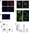
Fig. 1.
Fig. 1.
Tim-4 undergoes dynamic localization, and its cytoplasmic tail is required during the phagocytosis of apoptotic cells. (A) Immunofluorescence images of rPMs from WT mice stained for Tim-4 (red) and nuclei (blue) without (Upper) or with (Lower) the addition of apoptotic cells (A.C.). Thymocytes are identified by their DAPI-stained nuclei with round morphology. The dotted line outlines the cell periphery. Arrows point to contact sites between macrophage and apoptotic cells with enriched Tim-4 signal. (B) 3D image of a macrophage in contact with apoptotic cells (stained blue with nuclear dye) revealing the concentrated Tim-4 (red) distribution to these contact sites (arrows). (C) Time-lapse images of a 293T cell expressing Tim-4-YFP. Apoptotic thymocytes were added at 0 min. Rings of YFP-rich structure (arrows) began to appear at 10 min after apoptotic cell addition. (D) Surface expression of full-length mTim-4-YFP (FL) and mTim-4ΔC-YFP (ΔC). 293T cells transfected with either construct were stained for cell surface Tim-4 without detergent permeabilization. Representative images are shown with or without the addition of apoptotic cells (A.C.). Arrow points to aggregate of surface Tim-4 signal. (E) FACS plot of YFP+ cells from 293T cells expressing YFP alone, full-length mTim-4-YFP (FL), or mTim-4ΔC-YFP (ΔC). After a 90-min incubation with DiI-labeled apoptotic thymocytes, extracellular thymocytes were stained for CD45. YFP+ cells that have phagocytosed apoptotic cells are DiI+ CD45 (gated). (F) Graph of phagocytic YFP+ cells relative to 293T cells expressing YFP alone. Each experiment was performed at least three times. Representative data are shown.
Fig. 2.
Fig. 2.
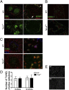
Tim-4 in the marginal zone and germinal center of the spleen is not required for apoptotic cell trapping and clearance or anti-SRBC antibody responses. (A) Tim-4 is expressed in and inside the marginal zone. Cryosections from WT and Tim-4−/− spleens 6 days after SRBC i.p. injection stained for peanut agglutinin (PNA) (green) and Tim-4 (red) (Left) or costained for the marginal metallophilic macrophage marker, MOMA-1 (green) and Tim-4 (red) (Right). The arrowhead indicates Tim-4+ MOMA-1+ cells in the marginal zone. Inside the marginal zone, Tim-4 can be detected in cells that are MOMA-1 (arrow). (B) Fluorescence images of a spleen section from animals injected with Thy1.1+ apoptotic thymocytes. Trapping of Thy1.1 apoptotic thymocytes (red) by MOMA-1 (green) is observed for both WT and Tim-4−/−. (C) Tingible bodies in spleens from WT and Tim-4−/− mice after primary SRBC challenge. Apoptotic bodies are shown in green by TUNEL staining. Tingible body macrophages are stained with CD68. Tim-4 is shown in blue. (D) Graph of average number of TUNEL+ cells per imaging field in the spleens of WT and Tim4−/− mice at 8 weeks and 6 months of age. Error bars: ±SEM. (E) Representative images of TUNEL+ (white dots) splenic sections from WT (Upper) and Tim4−/− (Lower) animals (n = 3). DAPI counterstain is shown (gray).
Fig. 3.
Fig. 3.
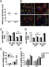
Unaltered immune function in Tim4−/− mice. (A) Change in ear thickness in WT and Tim4−/− mice DTH response induced by OVA (n = 6). (B) Structure of lymphoid follicles in spleens after SRBC challenge. Sections from Fig. 2A were stained with CD35 (green), B220 (red), and peanut agglutinin (PNA) (blue) for follicular DCs, B cells, and germinal center B cells, respectively. Serum IgM and IgG levels of anti-SRBCs after primary (C) and secondary (D) challenges are shown. The serum level of anti-dsDNA from aged (6–8 months old) WT and Tim4−/− mice (E) or from mice after each biweekly injection of apoptotic thymocytes (F) (n ≥ 7). The serum level of anti-dsDNA from the autoimmune-prone NZB/NZW F1 mouse strain is shown as a reference (gray bar in E). Error bar: ±SEM. Each immunization experiment was repeated once with n = 5.
Fig. 4.
Fig. 4.
Specific requirement of Tim-4 in the phagocytosis of apoptotic cells by rPMs. (A) rPMs fed with apoptotic thymocytes were stained for Thy1.2 (green), F4/80 (red), and DAPI (blue). Apoptotic cells internalized by macrophages appear as round nuclear-stained dots devoid of green staining inside red F4/80+ cells (arrowheads). Arrows depict extracellular apoptotic cells. (B) Quantification of internalized thymocytes per 100 macrophages relative to WT. (CH) In vivo phagocytosis by rPMs. DiI-labeled apoptotic thymocytes were injected i.p. into WT or Tim-4−/− mice, and peritoneal cells were harvested at 30 min and stained with Thy1.2 to identify extracellular thymocytes. FACS plots from peritoneal cells from animals injected with apoptotic cells (C) and the corresponding histogram of DiI intensity in the F4/80+ population (D) are shown. Filled gray curves are macrophages without apoptotic cells. F4/80+ macrophages that were DiI+ Thy1.2 were scored as phagocytic. (E) Percent of phagocytic macrophages that engulfed apoptotic cells were expressed relative to WT. (FH) Phagocytosis of apoptotic thymocytes by thioglycollate-elicited peritoneal macrophages from WT and Tim-4−/− mice. (F) FACS plots of DiI (Left) and DiI and Thy1.2 (Right) within the F4/80+ population. (G) Histogram of DiI intensity in the F4/80+ population. The filled gray curve represents macrophages without apoptotic thymocytes. (H) Graph of phagocytic thioglycollate-elicited peritoneal macrophages relative to WT. Each experiment was repeated at least twice.
Fig. 5.
Fig. 5.
Tim-4 deficiency causes increased peritoneal cell number. Peritoneal cells from WT and Tim-4−/− mice were collected by lavage (n = 3). (A) Total number of peritoneal cells from WT and Tim-4−/− mice. (B) Number of cells in lymphoid and myeloid compartments was determined by gating based on forward and side scatter. Error bar: ±SEM.
Fig. 6.
Fig. 6.
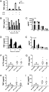
Tim-4−/− rPMs produce elevated basal TNF-α and exhibit dampened TNF-α production when stimulated with LPS. (A) TNF-α level in supernatant from rPMs cultured for 4 h without or with 100 ng/mL LPS in the absence or presence of apoptotic thymocytes (A.C.) measured by ELISA. The graph shows representative data from two independent experiments. TNF-α concentration in supernatant from rPMs cultured in the presence (B) or absence (D) of 100 ng/mL LPS for the indicated time. Levels of TNF-α, IL-6, IL-10, and IL-12(p40) in supernatant of resident peritoneal cell culture at 4 h in the presence (C) or absence (E) of LPS. Shown are representative data from three independent experiments. (FI) Levels of IL-17, IFN-γ, TNF-α, and keratinocyte-derived chemokine (KC) in peritoneal lavage from WT and Tim4−/− mice injected i.p. with PBS or E. coli. *, measurement below detection limit. Error bar: ±SEM.

References

    1. McIntire JJ, et al. Identification of Tapr (an airway hyperreactivity regulatory locus) and the linked Tim gene family. Nat Immunol. 2001;2:1109–1116. - PubMed
    1. Kuchroo VK, Dardalhon V, Xiao S, Anderson AC. New roles for TIM family members in immune regulation. Nature Review Immunology. 2008;8:577–580. - PubMed
    1. Meyers JH, et al. TIM-4 is the ligand for TIM-1, and the TIM-1-TIM-4 interaction regulates T cell proliferation. Nat Immunol. 2005;6:455–464. - PubMed
    1. Miyanishi M, et al. Identification of Tim4 as a phosphatidylserine receptor. Nature. 2007;450:435–439. - PubMed
    1. Kobayashi N, et al. TIM-1 and TIM-4 glycoproteins bind phosphatidylserine and mediate uptake of apoptotic cells. Immunity. 2007;27:927–940. - PMC - PubMed

MeSH terms

LinkOut - more resources