. 2010 May 11;107(19):8683-8.
doi: 10.1073/pnas.1003680107. Epub 2010 Apr 26.

Alternate protein kinase A activity identifies a unique population of stromal cells in adult bone

Affiliations

Alternate protein kinase A activity identifies a unique population of stromal cells in adult bone

Kit Man Tsang et al. Proc Natl Acad Sci U S A. .

Abstract

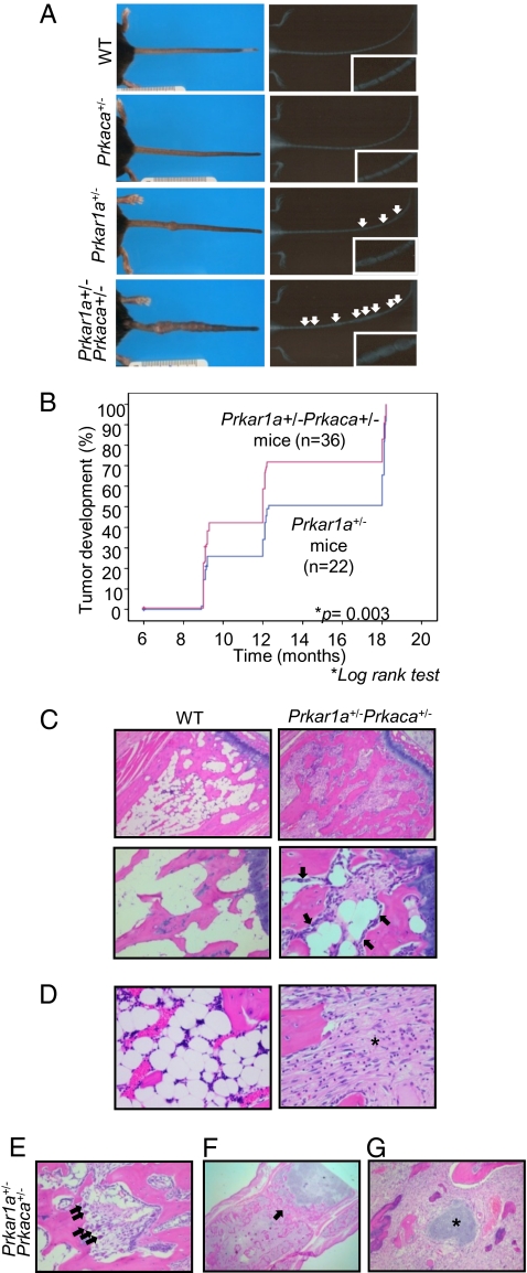
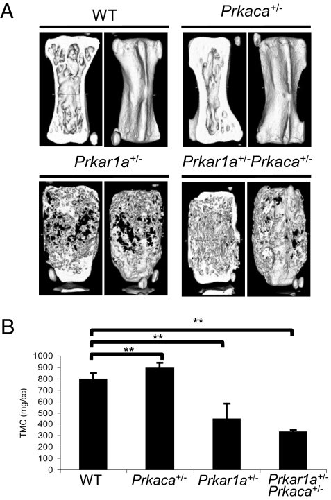
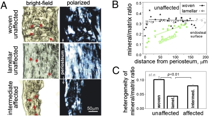
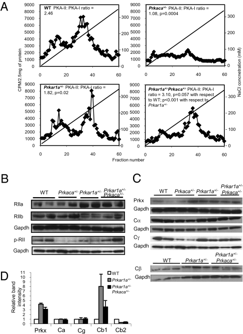
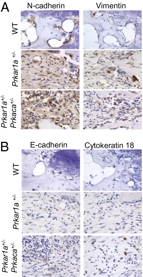
A population of stromal cells that retains osteogenic capacity in adult bone (adult bone stromal cells or aBSCs) exists and is under intense investigation. Mice heterozygous for a null allele of prkar1a (Prkar1a(+/-)), the primary receptor for cyclic adenosine monophosphate (cAMP) and regulator of protein kinase A (PKA) activity, developed bone lesions that were derived from cAMP-responsive osteogenic cells and resembled fibrous dysplasia (FD). Prkar1a(+/-) mice were crossed with mice that were heterozygous for catalytic subunit Calpha (Prkaca(+/-)), the main PKA activity-mediating molecule, to generate a mouse model with double heterozygosity for prkar1a and prkaca (Prkar1a(+/-)Prkaca(+/-)). Unexpectedly, Prkar1a(+/-)Prkaca(+/-) mice developed a greater number of osseous lesions starting at 3 months of age that varied from the rare chondromas in the long bones and the ubiquitous osteochondrodysplasia of vertebral bodies to the occasional sarcoma in older animals. Cells from these lesions originated from an area proximal to the growth plate, expressed osteogenic cell markers, and showed higher PKA activity that was mostly type II (PKA-II) mediated by an alternate pattern of catalytic subunit expression. Gene expression profiling confirmed a preosteoblastic nature for these cells but also showed a signature that was indicative of mesenchymal-to-epithelial transition and increased Wnt signaling. These studies show that a specific subpopulation of aBSCs can be stimulated in adult bone by alternate PKA and catalytic subunit activity; abnormal proliferation of these cells leads to skeletal lesions that have similarities to human FD and bone tumors.


Conflict of interest statement

The authors declare no conflict of interest.

Figures

Fig. 1.
Fig. 1.
Development of bone lesions along the tail of Prkar1a+/− and Prkar1a+/−Prkaca+/− mice. (A Left) comparison of tails from WT, Prkaca+/−, Prkar1a+/−, and Prkar1a+/−Prkaca+/− mice at 12 months old. (A Right) X-ray radiographs. White arrows point to the lesions. (B) Kaplan–Meier curve shows the number of tail masses found in various ages of Prkar1a+/− and Prkar1a+/−Prkaca+/− mice. (CG) Hematoxylin and eosin (H&E) staining of longitudinal sections of WT bones and Prkar1a+/−Prkaca+/− bone lesions. In C, black arrows denote the presence of mature osteoblasts lining along the trabecular bone. (Original magnification: C Upper, ×10; Lower, original magnification, ×20 . In D, the asterisk denotes the bone marrow space filled with fibroblastoid cells. (Original magnification, ×40). In E, black arrows denote the presence of osteoclasts. (Original magnification: ×20.) In F, the black arrow denotes the destroyed joint space. (Original magnification: ×2.) In G, the asterisk denotes the presence of cartilage island within the fibroblasts. (Original magnification: ×20.)
Fig. 2.
Fig. 2.
Undermineralization of bone in both Prkar1a+/− and Prkar1a+/−Prkaca+/− mice. (A) μCT images of caudal vertebra from WT, Prkaca+/−, Prkar1a+/− , and Prkar1a+/−Prkaca+/− mice at the age of 12 months. (B) Average of tissue mineral content (TMC) measurement of three caudal vertebrae from WT, Prkaca+/−, Prkar1a+/−, and Prkar1a+/−Prkaca+/− mice at 12 months old. **, P < 0.01. Error bars represent means ± SD.
Fig. 3.
Fig. 3.
Structure and mineralization of cortical bone in adjacent affected and unaffected caudal vertebrae. (A) Brightfield and polarized images of unaffected and affected caudal vertebrae. Well organized, lamellar/fine-fibered bone was indicated by well oriented spindle-shaped osteocyte lacunae (arrows) and more uniform polarized images due to regular collagen fiber orientation. Woven bone was indicated by irregular-shaped, disoriented lacunae and patchy appearance of polarized images due to irregular fiber orientation. (B) Even (−0.1 ± 0.2 mm−1 slope) mineral/matrix ratio across the cortical layer (the intensity ratio of mineral PO4 to organic CH Raman peaks) is characteristic of well mineralized, mature bone in unaffected vertebrae. Gradually increasing mineral/matrix ratio from periosteal to endosteal surface [+0.8 ± 0.2 (SD) mm−1 slope, P < 0.003] indicates lagging mineralization characteristic of rapidly growing, immature bone in affected vertebrae. (C) High mineralization heterogeneity (coefficient of variation for the mineral/matrix ratio) in all cortical regions of affected vertebrae is also consistent with rapid formation of immature bone.
Fig. 4.
Fig. 4.
Increased PKA-II complex, type II regulatory subunit and catalytic subunit β1, and Prkx in Prkar1a+/−Prkaca+/− mice bone tumors. (A) DEAE-chromatography of PKA isozymes in tail tissues of WT and Prkaca+/− mice and tail lesions of Prkar1a+/− and Prkar1a+/−Prkaca+/− mice. PKA-II to PKA-I ratio was calculated from averaging the intensities of 10 fractions within the peaks. Note that tail lesions of Prkar1a+/−Prkaca+/− mice had the highest PKA-II to PKA-I ratio (n = 3). (B) Western blot analysis on RIIα, RIIβ, and phosphorylated form of RII in WT, Prkaca+/−, Prkar1a+/−, and Prkar1a+/−Prkaca+/− mice at 1 year of age, showing the up-regulation of RII subunits in bone lesions and increase in phosphorylated form of RII in Prkar1a+/−Prkaca+/− tumors. (C) Western blot analysis on different PKA catalytic subunits of WT, Prkaca+/−, Prkar1a+/−, and Prkar1a+/−Prkaca+/− mice at 1 year of age. (D) Relative quantification of Prkx, Cα, Cγ, Cβ1, and Cβ2 protein in bone lesions against WT normal bone.
Fig. 5.
Fig. 5.
Expression of mesenchymal proteins in bone lesions from Prkar1a+/− and Prkar1a+/−Prkaca+/− mice and epithelial markers in Prkar1a+/−Prkaca+/− bone tumors that confirms the mesenchymal-to-epithelial gene signature in lesions from Prkar1a+/−Prkaca+/− mice. (A) Immunohistochemistry for n-cadherin and vimentin, mesenchymal proteins, is increased in all animals of PKA defects. (B) Immunohistochemistry for e-cadherin and cytokeratin 18, epithelial proteins, is increased in double heterozygote animals only.

Similar articles

Cited by

References

    1. Bossis I, Stratakis CA. Minireview: PRKAR1A: normal and abnormal functions. Endocrinology. 2004;145:5452–5458. - PubMed
    1. Skalhegg BS, Tasken K. Specificity in the cAMP/PKA signaling pathway. Differential expression,regulation, and subcellular localization of subunits of PKA. Front Biosci. 2000;5:D678–D693. - PubMed
    1. Gamm DM, Baude EJ, Uhler MD. The major catalytic subunit isoforms of cAMP-dependent protein kinase have distinct biochemical properties in vitro and in vivo. J Biol Chem. 1996;271:15736–15742. - PubMed
    1. McKnight GS, et al. Analysis of the cAMP-dependent protein kinase system using molecular genetic approaches. Recent Prog Horm Res. 1988;44:307–335. - PubMed
    1. Kirschner LS, et al. A mouse model for the Carney complex tumor syndrome develops neoplasia in cyclic AMP-responsive tissues. Cancer Res. 2005;65:4506–4514. - PubMed

Publication types

MeSH terms

Substances

Associated data