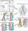
Structure elucidation of dimeric transmembrane domains of bitopic proteins
- PMID: 20421711
- PMCID: PMC2900626
- DOI: 10.4161/cam.4.2.11930
Structure elucidation of dimeric transmembrane domains of bitopic proteins
Abstract
The interaction between transmembrane helices is of great interest because it directly determines biological activity of a membrane protein. Either destroying or enhancing such interactions can result in many diseases related to dysfunction of different tissues in human body. One much studied form of membrane proteins known as bitopic protein is a dimer containing two membrane-spanning helices associating laterally. Establishing structure-function relationship as well as rational design of new types of drugs targeting membrane proteins requires precise structural information about this class of objects. At present time, to investigate spatial structure and internal dynamics of such transmembrane helical dimers, several strategies were developed based mainly on a combination of NMR spectroscopy, optical spectroscopy, protein engineering and molecular modeling. These approaches were successfully applied to homo- and heterodimeric transmembrane fragments of several bitopic proteins, which play important roles in normal and in pathological conditions of human organism.
Figures


Similar articles
-
Structural and thermodynamic insight into the process of "weak" dimerization of the ErbB4 transmembrane domain by solution NMR.Biochim Biophys Acta. 2012 Sep;1818(9):2158-70. doi: 10.1016/j.bbamem.2012.05.001. Epub 2012 May 8. Biochim Biophys Acta. 2012. PMID: 22579757
-
Structural aspects of oligomerization taking place between the transmembrane alpha-helices of bitopic membrane proteins.Biochim Biophys Acta. 2002 Oct 11;1565(2):347-63. doi: 10.1016/s0005-2736(02)00580-1. Biochim Biophys Acta. 2002. PMID: 12409206 Review.
-
Helix-helix interactions in membrane domains of bitopic proteins: Specificity and role of lipid environment.Biochim Biophys Acta Biomembr. 2017 Apr;1859(4):561-576. doi: 10.1016/j.bbamem.2016.10.024. Epub 2016 Nov 22. Biochim Biophys Acta Biomembr. 2017. PMID: 27884807 Review.
-
TMDIM: an improved algorithm for the structure prediction of transmembrane domains of bitopic dimers.J Comput Aided Mol Des. 2017 Sep;31(9):855-865. doi: 10.1007/s10822-017-0047-0. Epub 2017 Sep 1. J Comput Aided Mol Des. 2017. PMID: 28864946
-
How important are transmembrane helices of bitopic membrane proteins?Biochim Biophys Acta. 2007 Mar;1768(3):387-92. doi: 10.1016/j.bbamem.2006.11.019. Epub 2006 Dec 8. Biochim Biophys Acta. 2007. PMID: 17258687
Cited by
-
Peripheral membrane associations of matrix metalloproteinases.Biochim Biophys Acta Mol Cell Res. 2017 Nov;1864(11 Pt A):1964-1973. doi: 10.1016/j.bbamcr.2017.04.013. Epub 2017 Apr 23. Biochim Biophys Acta Mol Cell Res. 2017. PMID: 28442379 Free PMC article. Review.
-
New Insights into Molecular Organization of Human Neuraminidase-1: Transmembrane Topology and Dimerization Ability.Sci Rep. 2016 Dec 5;6:38363. doi: 10.1038/srep38363. Sci Rep. 2016. PMID: 27917893 Free PMC article.
-
Structural and Functional Insights into the Transmembrane Domain Association of Eph Receptors.Int J Mol Sci. 2021 Aug 10;22(16):8593. doi: 10.3390/ijms22168593. Int J Mol Sci. 2021. PMID: 34445298 Free PMC article. Review.
-
Inconspicuous Yet Indispensable: The Coronavirus Spike Transmembrane Domain.Int J Mol Sci. 2023 Nov 16;24(22):16421. doi: 10.3390/ijms242216421. Int J Mol Sci. 2023. PMID: 38003610 Free PMC article. Review.
-
When detergent meets bilayer: birth and coming of age of lipid bicelles.Prog Nucl Magn Reson Spectrosc. 2013 Feb;69:1-22. doi: 10.1016/j.pnmrs.2013.01.001. Epub 2013 Jan 23. Prog Nucl Magn Reson Spectrosc. 2013. PMID: 23465641 Free PMC article. Review. No abstract available.
References
-
- Ubarretxena-Belandia I, Engelman DM. Helical membrane proteins: diversity of functions in the context of simple architecture. Curr Opin Struct Biol. 2001;11:370–376. - PubMed
-
- Schlessinger J. Cell signaling by receptor tyrosine kinases. Cell. 2000;281:211–225. - PubMed
-
- Moriki T, Maruyama H, Maruyama IN. Activation of preformed EGF receptor dimers by ligand-induced rotation of the transmembrane domain. J Mol Biol. 2001;311:1011–1026. - PubMed
Publication types
MeSH terms
Substances
LinkOut - more resources
Full Text Sources
