

Memory CD8+ T cells from naturally acquired primary dengue virus infection are highly cross-reactive
- PMID: 20421879
- PMCID: PMC2929403
- DOI: 10.1038/icb.2010.61
Memory CD8+ T cells from naturally acquired primary dengue virus infection are highly cross-reactive
Abstract
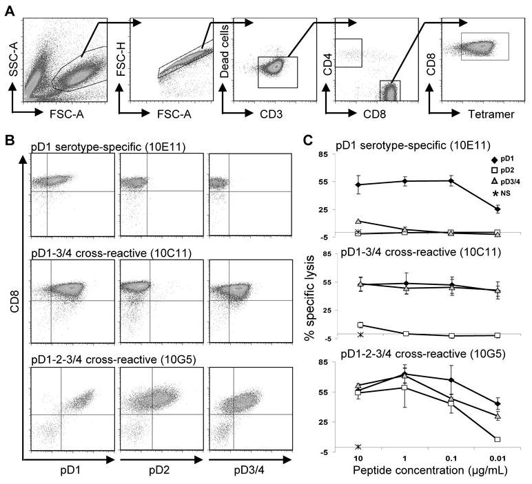
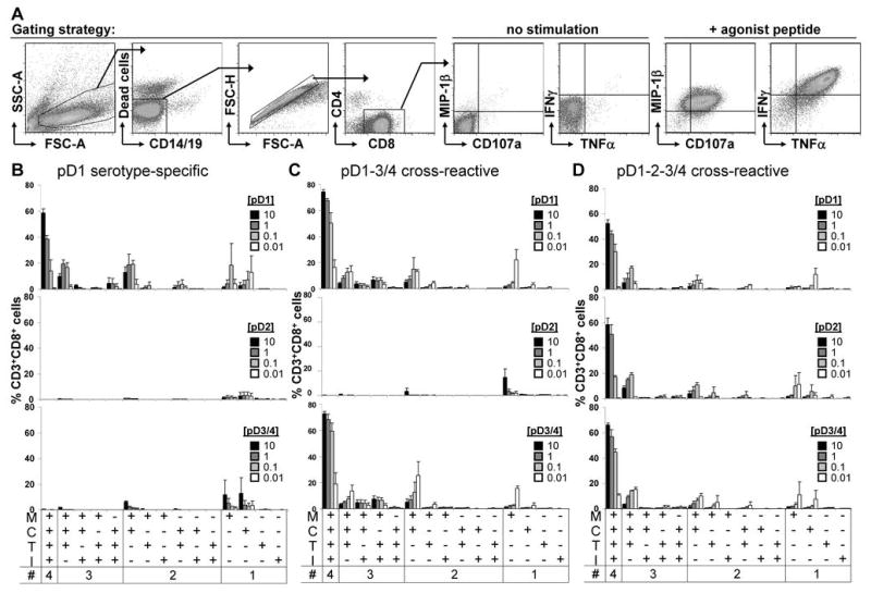
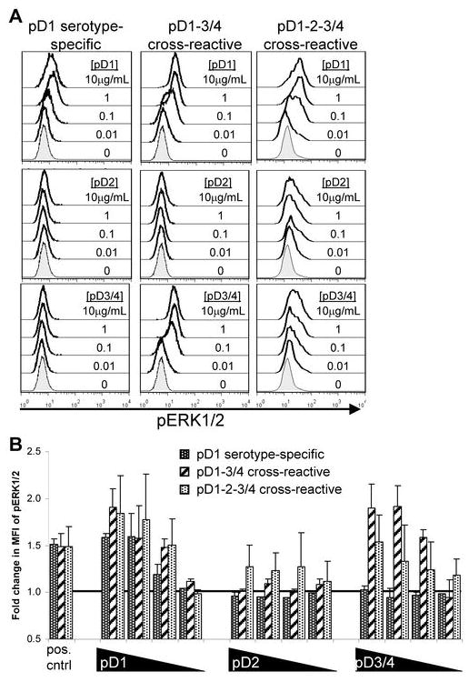
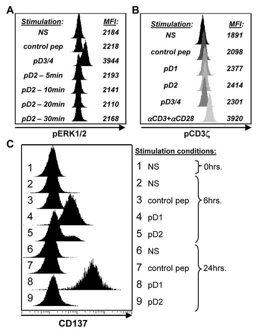
Cross-reactive memory T cells induced by primary infection with one of the four serotypes of dengue virus (DENV) are hypothesized to have an immunopathological function in secondary heterologous DENV infection. To define the T-cell response to heterologous serotypes, we isolated HLA-A(*)1101-restricted epitope-specific CD8(+) T-cell lines from primary DENV-immune donors. Cell lines exhibited marked cross-reactivity toward peptide variants representing the four DENV serotypes in tetramer binding and functional assays. Many clones responded similarly to homologous and heterologous serotypes with striking cross-reactivity between the DENV-1 and DENV-3 epitope variants. In vitro-stimulated T-cell lines consistently revealed a hierarchical induction of MIP-1β>degranulation>tumor necrosis factor α (TNFα)>interferon-γ (IFNγ), which depended on the concentration of agonistic peptide. Phosphoflow assays showed peptide dose-dependent phosphorylation of ERK1/2, which correlated with cytolysis, degranulation, and induction of TNFα and IFNγ, but not MIP-1β production. This is the first study to show significant DENV serotype-cross-reactivity of CD8(+) T cells after naturally acquired primary infection. We also show qualitatively different T-cell receptor signaling after stimulation with homologous and heterologous peptides. Our data support a model whereby the order of sequential DENV infections influences the immune response to secondary heterologous DENV infection, contributing to varying disease outcomes.
Figures
References
-
- Pinheiro FP, Corber SJ. Global situation of dengue and dengue haemorrhagic fever, and its emergence in the Americas. World Health Stat Q. 1997;50:161–169. - PubMed
-
- Wilder-Smith A, Gubler DJ. Geographic expansion of dengue: the impact of international travel. Med Clin North Am. 2008;92:1377–1390. x. - PubMed
-
- Thein S, Aung MM, Shwe TN, Aye M, Zaw A, Aye K, et al. Risk factors in dengue shock syndrome. Am J Trop Med Hyg. 1997;56:566–572. - PubMed
-
- Guzman MG, Kouri GP, Bravo J, Soler M, Vazquez S, Morier L. Dengue hemorrhagic fever in Cuba, 1981: a retrospective seroepidemiologic study. Am J Trop Med Hyg. 1990;42:179–184. - PubMed
-
- Burke DS, Nisalak A, Johnson DE, Scott RM. A prospective study of dengue infections in Bangkok. Am J Trop Med Hyg. 1988;38:172–180. - PubMed
Publication types
MeSH terms
Substances
Grants and funding
LinkOut - more resources
Full Text Sources
Other Literature Sources
Medical
Research Materials
Miscellaneous
