

Novel snail1 target proteins in human colon cancer identified by proteomic analysis
- PMID: 20421926
- PMCID: PMC2857666
- DOI: 10.1371/journal.pone.0010221
Novel snail1 target proteins in human colon cancer identified by proteomic analysis
Abstract
Background: The transcription factor Snail1 induces epithelial-to-mesenchymal transition (EMT), a process responsible for the acquisition of invasiveness during tumorigenesis. Several transcriptomic studies have reported Snail1-regulated genes in different cell types, many of them involved in cell adhesion. However, only a few studies have used proteomics as a tool for the characterization of proteins mediating EMT.
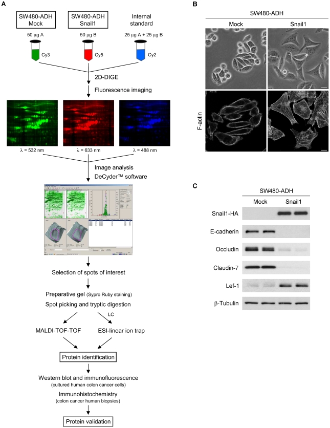
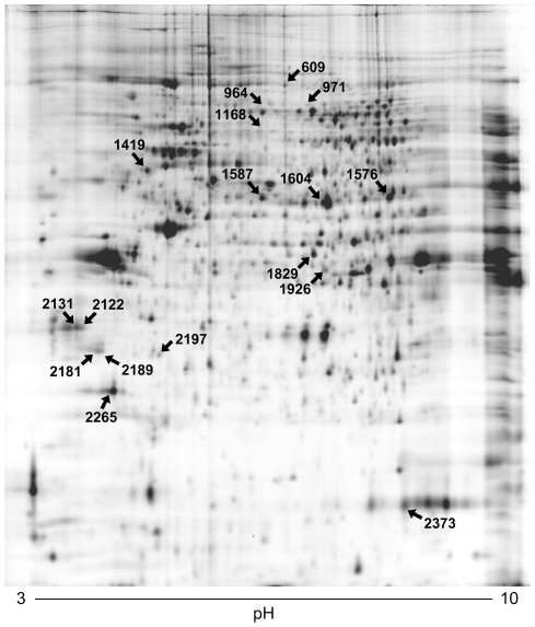
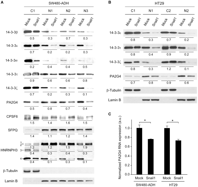
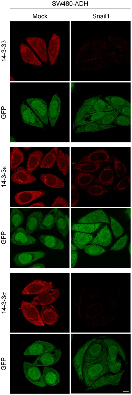
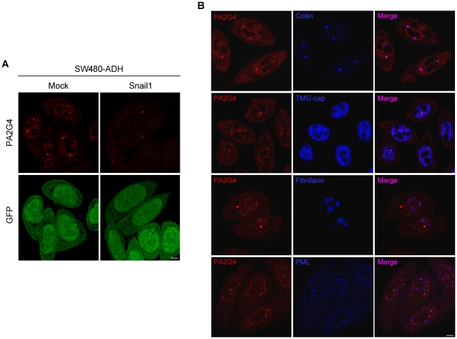
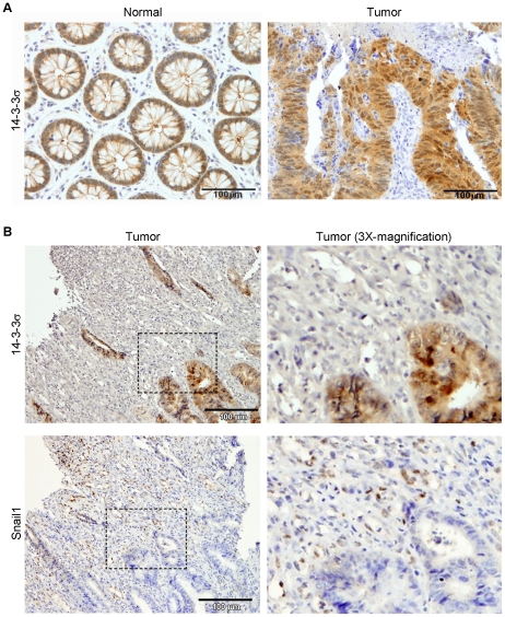
Methodology/principal findings: We identified by proteomic analysis using 2D-DIGE electrophoresis combined with MALDI-TOF-TOF and ESI-linear ion trap mass spectrometry a number of proteins with variable functions whose expression is modulated by Snail1 in SW480-ADH human colon cancer cells. Validation was performed by Western blot and immunofluorescence analyses. Snail1 repressed several members of the 14-3-3 family of phosphoserine/phosphothreonine binding proteins and also the expression of the Proliferation-associated protein 2G4 (PA2G4) that was mainly localized at the nuclear Cajal bodies. In contrast, the expression of two proteins involved in RNA processing, the Cleavage and polyadenylation specificity factor subunit 6 (CPSF6) and the Splicing factor proline/glutamine-rich (SFPQ), was higher in Snail1-expressing cells than in controls. The regulation of 14-3-3epsilon, 14-3-3tau, 14-3-3zeta and PA2G4 by Snail1 was reproduced in HT29 colon cancer cells. In addition, we found an inverse correlation between 14-3-3sigma and Snail1 expression in human colorectal tumors.
Conclusions/significance: We have identified a set of novel Snail1 target proteins in colon cancer that expand the cellular processes affected by Snail1 and thus its relevance for cell function and phenotype.
Conflict of interest statement
Figures
Similar articles
-
Differential expression of proteins in response to ceramide-mediated stress signal in colon cancer cells by 2-D gel electrophoresis and MALDI-TOF-MS.J Proteome Res. 2005 May-Jun;4(3):870-80. doi: 10.1021/pr050006t. J Proteome Res. 2005. PMID: 15952734
-
Four and a half LIM protein 2 (FHL2) negatively regulates the transcription of E-cadherin through interaction with Snail1.Eur J Cancer. 2011 Jan;47(1):121-30. doi: 10.1016/j.ejca.2010.07.045. Eur J Cancer. 2011. PMID: 20801642
-
Genome-wide mapping of DNA-binding sites identifies stemness-related genes as directly repressed targets of SNAIL1 in colorectal cancer cells.Oncogene. 2019 Oct;38(40):6647-6661. doi: 10.1038/s41388-019-0905-4. Epub 2019 Aug 7. Oncogene. 2019. PMID: 31391555
-
Proteomic analysis of transcription factor interactions in myeloid stem cell development and leukaemia.Expert Opin Ther Targets. 2002 Aug;6(4):491-5. doi: 10.1517/14728222.6.4.491. Expert Opin Ther Targets. 2002. PMID: 12223063 Review.
-
Dual role of Snail1 as transcriptional repressor and activator.Biochim Biophys Acta Rev Cancer. 2024 Jan;1879(1):189037. doi: 10.1016/j.bbcan.2023.189037. Epub 2023 Dec 2. Biochim Biophys Acta Rev Cancer. 2024. PMID: 38043804 Review.
Cited by
-
Proteogenomic analysis reveals unanticipated adaptations of colorectal tumor cells to deficiencies in DNA mismatch repair.Cancer Res. 2014 Jan 1;74(1):387-97. doi: 10.1158/0008-5472.CAN-13-2488. Epub 2013 Nov 18. Cancer Res. 2014. PMID: 24247723 Free PMC article.
-
The application of the open pharmacological concepts triple store (open PHACTS) to support drug discovery research.PLoS One. 2014 Dec 18;9(12):e115460. doi: 10.1371/journal.pone.0115460. eCollection 2014. PLoS One. 2014. PMID: 25522365 Free PMC article.
-
Epithelial mesenchymal transition and tumor budding in aggressive colorectal cancer: tumor budding as oncotarget.Oncotarget. 2010 Nov;1(7):651-61. doi: 10.18632/oncotarget.199. Oncotarget. 2010. PMID: 21317460 Free PMC article. Review.
-
HIV-1-induced translocation of CPSF6 to biomolecular condensates.Trends Microbiol. 2024 Aug;32(8):781-790. doi: 10.1016/j.tim.2024.01.001. Epub 2024 Jan 23. Trends Microbiol. 2024. PMID: 38267295 Free PMC article. Review.
-
The role of Snail1 transcription factor in colorectal cancer progression and metastasis.Contemp Oncol (Pozn). 2015;19(4):265-70. doi: 10.5114/wo.2014.42173. Epub 2015 Sep 28. Contemp Oncol (Pozn). 2015. PMID: 26557772 Free PMC article. Review.
References
-
- Thiery JP, Acloque H, Huang RYJ, Nieto MA. Epithelial-mesenchymal transitions in development and disease. Cell. 2009;139:871–890. - PubMed
-
- Peinado H, Olmeda D, Cano A. Snail, Zeb and bHLH factors in tumour progression: an alliance against the epithelial phenotype? Nat Rev Cancer. 2007;7:415–428. - PubMed
-
- Moreno-Bueno G, Cubillo E, Sarrió D, Peinado H, Rodríguez-Pinilla SM, et al. Genetic profiling of epithelial cells expressing E-cadherin repressors reveals a distinct role for Snail, Slug, and E47 factors in epithelial-mesenchymal transition. Cancer Res. 2006;66:9543–9556. - PubMed
-
- Batlle E, Sancho E, Francí C, Domínguez D, Monfar M, et al. The transcription factor Snail is a repressor of E-cadherin gene expression in epithelial tumour cells. Nat Cell Biol. 2000;2:84–89. - PubMed
