Comparative Study
. 2010 Apr 20;4(4):e666.
doi: 10.1371/journal.pntd.0000666.

Mosquito transcriptome profiles and filarial worm susceptibility in Armigeres subalbatus

Affiliations
Comparative Study

Mosquito transcriptome profiles and filarial worm susceptibility in Armigeres subalbatus

Matthew T Aliota et al. PLoS Negl Trop Dis. .

Abstract

Background: Armigeres subalbatus is a natural vector of the filarial worm Brugia pahangi, but it kills Brugia malayi microfilariae by melanotic encapsulation. Because B. malayi and B. pahangi are morphologically and biologically similar, comparing Ar. subalbatus-B. pahangi susceptibility and Ar. subalbatus-B. malayi refractoriness could provide significant insight into recognition mechanisms required to mount an effective anti-filarial worm immune response in the mosquito, as well as provide considerable detail into the molecular components involved in vector competence. Previously, we assessed the transcriptional response of Ar. subalbatus to B. malayi, and now we report transcriptome profiling studies of Ar. subalbatus in relation to filarial worm infection to provide information on the molecular components involved in B. pahangi susceptibility.

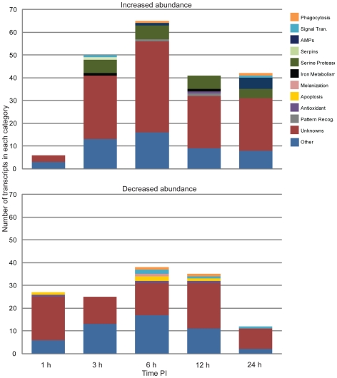
Methodology/principal findings: Utilizing microarrays, comparisons were made between mosquitoes exposed to B. pahangi, B. malayi, and uninfected bloodmeals. The time course chosen facilitated an examination of key events in the development of the parasite, beginning with the very start of filarial worm infection and spanning to well after parasites had developed to the infective stage in the mosquito. At 1, 3, 6, 12, 24 h post infection and 2-3, 5-6, 8-9, and 13-14 days post challenge there were 31, 75, 113, 76, 54, 5, 3, 13, and 2 detectable transcripts, respectively, with significant differences in transcript abundance (increase or decrease) as a result of parasite development.

Conclusions/significance: Herein, we demonstrate that filarial worm susceptibility in a laboratory strain of the natural vector Ar. subalbatus involves many factors of both known and unknown function that most likely are associated with filarial worm penetration through the midgut, invasion into thoracic muscle cells, and maintenance of homeostasis in the hemolymph environment. The data show that there are distinct and separate transcriptional patterns associated with filarial worm susceptibility as compared to refractoriness, and that an infection response in Ar. subalbatus can differ significantly from that observed in Ae. aegypti, a common laboratory model.


Conflict of interest statement

The authors have declared that no competing interests exist.

Figures

Figure 1
Figure 1. Transcriptional changes in Armigeres subalbatus following a Brugia pahangi-infected bloodmeal.
The bar graph represents the number of significantly changed transcripts over the course of experimentation. Volcano plots were used to create working gene lists to identify differentially expressed transcripts at each time point. At 1, 3, 6, 12, 24 h post infection and 2–3, 5–6, 8–9, and 13–14 d post challenge there were 29, 75, 103, 76, 54, 5, 3, 13, and 2 detectable transcripts, respectively, with significantly different transcriptional behavior (increased or decreased transcript abundance at a 95% confidence interval over two-fold values) as a result of parasite development. The sample groups chosen are defined by the time post ingestion of the bloodmeal and represent significantly different stages of parasite development. The bar graph represents groups of mosquitoes that included those exposed to an infective bloodmeal containing B. pahangi mf (∼15–60 mf/20 µl blood) and those exposed to a bloodmeal without parasites.
Figure 2
Figure 2. Functional composition of transcripts significantly affected by parasite infection.
The categories are based on abundant and immunity-related EST clusters observed from Armigeres subalbatus cDNA libraries. Transcripts with a detectable increase in abundance (top) and with a detectable decrease in abundance (bottom) 1, 3, 6, 12, and 24 hours after exposure to a Brugia pahangi infective blood meal.
Figure 3
Figure 3. Transcriptional changes in Armigeres subalbatus when comparing a Brugia pahangi- vs. a Brugia malayi-infected bloodmeal.
The bar graph represents the number of genes with a significant fold difference between B. pahangi- or B. malayi-exposed mosquitoes. Volcano plots were used to create working gene lists to identify transcripts associated with B. pahangi development (left) vs. B. malayi resistance (right). At 1, 6, 12, 24 h post infection and 2–3 d post challenge there were 10, 14, 15, 27, and 4 detectable transcripts, respectively, more associated with B. pahangi infection. Following the same time course, there were 63, 20, 57, 81, and 6 detectable transcripts, respectively, more associated with B. malayi resistance. The bar graph represents groups of mosquitoes that included those exposed to blood containing B. pahangi mf and those exposed to blood containing B. malayi mf. Significant increases or decreases in transcript abundance cannot be delineated.
Figure 4
Figure 4. Functional composition of transcripts associated with Brugia pahangi infection or Brugia malayi resistance.
The categories are based on abundant and immunity-related EST clusters observed from Armigeres subalbatus cDNA libraries. Transcripts associated with B. pahangi infection (top). Transcripts associated with B. malayi resistance (bottom).
Figure 5
Figure 5. Development of Brugia pahangi in the mosquito thorax.
At 6 days post-infection parasites (e.g., arrows) are developing in the thoracic musculature and are oriented parallel to the myofibers (A). At 9 days post-infection parasites exit the myofibers (B) and migrate to the cervix and head region (C; 14 days post-injection). *, thoracic cuticle; #, cervical cuticle; scale bar, 100 µm.
Figure 6
Figure 6. Comparative histological imaging of thoracic indirect flight muscles after Brugia pahangi-infected and uninfected bloodmeals.
Aside from occasional host-derived tissue pooling between the worms and the myofibrils (A, A inset), no obvious pathology associated with infection was observed at 6 days (A, B), 9 days (C, D) or 14 days (E, F) after mosquitoes received Brugia pahangi-infected (A, C, E) and uninfected (B, D, F) bloodmeals. B. pahangi, yellow arrows; host tissue pooling, black arrow; *, fat body; A–F scale bar, 100 µm; A inset scale bar, 25 µm).

Similar articles

Cited by

References

    1. Mathers CD, Ezzati M, Lopez AD. Measuring the burden of neglected tropical diseases: the global burden of disease framework. PLoS Negl Trop Dis. 2007;1:e114. - PMC - PubMed
    1. Molyneux DH. Control of human parasitic diseases: Context and overview. Adv Parasitol. 2006;61:1–45. - PubMed
    1. Muturi EJ, Jacob BG, Kim CH, Mbogo CM, Novak RJ. Are coinfections of malaria and filariasis of any epidemiological significance? Parasitol Res. 2008;102:175–181. - PubMed
    1. Carton Y, Nappi AJ, Poirie M. Genetics of anti-parasite resistance in invertebrates. Dev Comp Immunol. 2005;29:9–32. - PubMed
    1. Pichon G. Limitation and facilitation in the vectors and other aspects of the dynamics of filarial transmission: the need for vector control against Anopheles-transmitted filariasis. Ann Trop Med Parasitol. 2002;96(Suppl 2):S143–152. - PubMed

Publication types