. 2010 Apr 22;6(4):e1000857.
doi: 10.1371/journal.ppat.1000857.

Proteolysis of human thrombin generates novel host defense peptides

Affiliations

Proteolysis of human thrombin generates novel host defense peptides

Praveen Papareddy et al. PLoS Pathog. .

Abstract

The coagulation system is characterized by the sequential and highly localized activation of a series of serine proteases, culminating in the conversion of fibrinogen into fibrin, and formation of a fibrin clot. Here we show that C-terminal peptides of thrombin, a key enzyme in the coagulation cascade, constitute a novel class of host defense peptides, released upon proteolysis of thrombin in vitro, and detected in human wounds in vivo. Under physiological conditions, these peptides exert antimicrobial effects against Gram-positive and Gram-negative bacteria, mediated by membrane lysis, as well as immunomodulatory functions, by inhibiting macrophage responses to bacterial lipopolysaccharide. In mice, they are protective against P. aeruginosa sepsis, as well as lipopolysaccharide-induced shock. Moreover, the thrombin-derived peptides exhibit helical structures upon binding to lipopolysaccharide and can also permeabilize liposomes, features typical of "classical" helical antimicrobial peptides. These findings provide a novel link between the coagulation system and host-defense peptides, two fundamental biological systems activated in response to injury and microbial invasion.


Conflict of interest statement

Drs. Schmidtchen and Malmsten have shares in DermaGen AB and XImmune AB, companies involved in the therapeutic development of peptides for topical and systemic use, respectively.

Figures

Figure 1
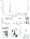
Figure 1. Generation of antimicrobial peptides by degradation of prothrombin and thrombin.
(A) Degradation of the proteins was performed at 37°C for the indicated time periods. RDA was performed in low-salt conditions using E. coli as test organism. Each 4 mm-diameter well was loaded with 6 µl of the solution (corresponding to 3.6 µg protein). The bar diagrams indicate the diameter of the zones of clearance obtained (in mm). The inset visualizes the results obtained with prothrombin. C, buffer; NE, neutrophil elastase only. LL-37 (100 µM) was included for comparison. (B) Intact prothrombin (PT) and thrombin (T), and cleavage products from the different incubations with neutrophil elastase (NE, indicated above) were analyzed by SDS-PAGE (16.5% Tris-Tricine gel). The gels are overloaded (12 µg) in order to visualize generation of fragments of low molecular masses. Rightmost two lanes show PT and T proteins at 2 µg.
Figure 2
Figure 2. Activities of peptides derived from prothrombin.
(A) Sequence of prothrombin and overlapping peptides (indicated by numbers). In addition to the regular overlapping peptides, peptide regions of high net charge, and/or content of predicted helical regions (Agadir; http://www.embl-heidelberg.de/Services/serrano/agadir/agadir-start.html) were selected. Peptides described in subsequent experiments are also indicated; GKY25, VFR17, and the major ∼11 kDa peptide (amino acids 527–622). (B) Overlapping peptides of prothrombin were analyzed for antimicrobial activities against E. coli. The inhibitory zones, relative hydrophobic moment (μHrel) as well as net charge of respective peptides (only active peptides are numbered) are indicated in the 3-D graph. Peptides showing antimicrobial activity are also indicated by red color in (A). For determination of antibacterial activities, E. coli (4×10⁶ cfu) was inoculated in 0.1% TSB agarose gel. Each 4 mm-diameter well was loaded with 6 µl of peptide (at 100 µM). The zones of clearance correspond to the inhibitory effect of each peptide after incubation at 37°C for 18–24 h (mean values are presented, n = 3). (C) Helical wheel representation of the C-terminal peptide VFR17. The amino acids are indicated. (D) LPS-binding activity of the prothrombin-derived peptide sequences. Peptides (5 µg) were applied to nitrocellulose membranes followed by incubation in PBS (containing 2% bovine serum albumin) with iodinated (125I)-LPS. Only peptides from the C-terminal part of prothrombin demonstrated significant binding to LPS. (E) Molecular model of thrombin. The peptides GKY25 (green-orange, indicated in Figure 2A) and VFR17 (orange, peptide 48 in Figure 2A) are indicated in the crystal structure of human thrombin (PDB code 1C5L). (F) Activities of prothrombin (PT), thrombin (T), GKY25 and VFR17 on E. coli ATCC 25922. In viable count assays GKY25 and VFR17 displayed significant antibacterial activities. 2×10⁶ cfu/ml of bacteria were incubated in 50 µl with proteins and peptides at a concentration of 3 and 6 µM, respectively.
Figure 3
Figure 3. Identification of antibacterial regions of thrombin and prothrombin.
(A) RP-HPLC separation of thrombin digested with neutrophil elastase. The bars indicate the antibacterial activity of the fractions in low (gray) as well as high salt conditions (black). Fraction 30 (lower left) contained two peaks of masses 2034.78 and 2270.88, perfectly matching the indicated sequences obtained after ESI-MS/MS analysis. Fraction 38 was analyzed by MALDI-MS, and subsequently by ESI-MS. The ESI-MS analysis identified a dominant mass of 11041 corresponding to the 96-amino acid long peptide N527-E622 (indicated in Figure 2A) with two intact disulphide bridges. A minor mass corresponding to V528-E622 was also detected by ESI-MS/MS. N- and C-terminal sequencing yelded NLPI and EGFQ, respectively. The rightmost insets illustrate the ∼11 kDa peptide analysed by SDS-PAGE and stained for protein (stain), or after immunoblot (blot), and below, the peptide (F38) was analyzed by gel-overlay for detection of antibacterial activity. The activity of F38 was identical to the major clearing zone generated by elastase-digested thrombin (T+NE). Right arrow indicates the position of clearing zone generated by the peptide GKY25. The gel was run top to bottom. Finally, peptides of fractions 20–21 were predicted using the FINDPEPT tool (www.expasy.org/tools/findpept.html) (Table S1). (B) Degradation of thrombin by neutrophil supernatants generates antibacterial activity in RDA (upper inset). RDA was performed in low-salt conditions. E. coli (4×10⁶ cfu) was used as test organism. Each 4 mm-diameter well was loaded with 6 µl of material (C, supernatant only; T, thrombin only; T+NS; thrombin incubated for 30 and 180 min respectively, with neutrophil supernatants). The digests were analysed by SDS-PAGE (16.5% Tris-Tricine gels) and immunoblotting with antibodies against VFR17 (lower panel). (C) Prothrombin was digested with the enzymes as indicated for 3 h, and analyzed by SDS-PAGE (16.5% Tris-Tricine gels) and immunoblotting using antibodies against VFR17 (NE, neutrophil elastase; CG, cathepsin G; PAE, P. aeruginosa elastase). (D) RDA results of prothrombin digested with cathepsin G (CG) and P. aeruginosa elastase (PAE) for different time periods. VFR17 and LL-37 (10 µM) are shown for comparison.
Figure 4
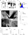
Figure 4. Thrombin-derived C-terminal peptides, their presence and antimicrobial effects ex vivo and in vivo.
(A) Fibrin clots were produced from human plasma and incubated with neutrophil elastase for the indicated time periods (Fibrin), or obtained from a patient with a venous, non-infected, chronic ulcer (PF), extracted, and analyzed by immunoblotting using polyclonal antibodies against the thrombin C-terminal peptide VFR17. (B) Human plasma, incubated with neutrophil elastase for the indicated time periods (Plasma, left panel), acute wound fluid (patients 1–2, AWF, middle panel), or wound fluid from patients with chronic ulcers (patients 1–6, CWF, right panel) was analysed by Western blot using polyclonal antibodies against the thrombin C-terminal peptide VFR17. (C) Flow cytometry analysis of binding of C-terminal thrombin epitopes to P. aeruginosa bacteria. Bacteria were incubated for 4 h with control plasma (P), human plasma depleted of prothrombin (DP), depleted plasma supplemented with the peptide GKY25, or, acute wound fluid (AWF). Binding of C-terminal epitopes to the bacteria was detected using primary antibodies against the C-terminal epitope VFR17 followed by addition of FITC-labeled secondary antibodies. (D) Visualization of binding and membrane damage by TCPs. P. aeruginosa bacteria were incubated ex vivo with human plasma (P), acute wound fluid (AWF), or wound fluid from a chronic leg ulcer (CWF), or visualized in vivo in fibrin slough (CWS) derived from a patient with a chronic ulcer infected by S. aureus. Arrows in P, AWF, and CWF point to damaged bacterial membranes. Coccoid bacteria (indicated by an arrow in CWS) show extensive binding of antibodies directed against the C-terminal peptide VFR17 (negative and positive bacterial controls, and additional material are found in Figure S3 and S4). (E) TCPs inhibit bacterial growth in human plasma. Control plasma (P), plasma depleted of prothrombin (DP), depleted plasma supplemented with either prothrombin (DP+PT), or GKY25 (DP+GKY25) (PT and GKY25 at 1.5 µM), or control AWF or depleted AWF (D-AWF), were inoculated with P. aeruginosa bacteria under similar conditions as in (C–D). The multiplication factors at various time points are given. After incubation, CFUs were determined by plating. Experiments were repeated three times and a representative experiment is shown. (F) The thrombin C-terminal peptide GKY25 significantly increases survival. Mice were injected i.p. with P. aeruginosa bacteria, followed by subcutaneous injection of GKY25 or buffer only, after 1 h. The injections were repeated after 24 hours. Treatment with the peptide significantly increased survival (n = 10 for controls and treated, p = 0.002). (G) GKY25 suppresses bacterial dissemination to the spleen and kidney. Mice were infected as above, GKY25 was administrated subcutaneously after 1 h, and the cfu of P. aeruginosa in spleen and kidney was determined after a time period of 8 h (n = 10 for controls and treated, P<0.05 for spleen and kidney. Horizontal line indicates median value).
Figure 5
Figure 5. LPS-binding and immunomodulatory role in vitro and in vivo of thrombin-derived C-terminal peptides.
(A) GKY25 binds heparin and LPS. 2 and 5 µg GKY25 were applied to nitrocellulose membranes. These membranes were then blocked in PBS (containing 2% bovine serum albumin) for 1 h at room temperature and incubated in PBS with iodinated (125I) heparin or LPS. Unlabeled heparin (6 mg/ml) (+) was added for competition of binding. LL-37 was used for comparison. The membranes were washed (3×10 min in PBS). A Bas 2000 radioimaging system (Fuji) was used for visualization of radioactivity. (B) GKY25 inhibits NO production. RAW264.7 macrophages were stimulated with LPS from E. coli and P. aeruginosa, in presence of GKY25 at the indicated concentrations. LL-37 is presented for comparison. (C) GKY25 significantly increases survival in LPS-induced shock. Mice were injected with LPS followed by intraperitoneal administration of GKY25 (200 µg). Survival was followed for 7 days. (n = 9 for controls, n = 10 for treated animals, P<0.001). (D) GKY25 attenuates proinflammatory cytokines. In a separate experiment, mice were sacrificed 6 h after i.p. injection of LPS followed by treatment with GKY25 (200 µg) or buffer, and the indicated cytokines were analyzed in blood (n = 9 for controls, n = 10 for treated animals, the P values for the respective cytokines are IL-6, 0.001; IFN-γ = 0.009; TNF, 0.001; IL-12p70, 0.001. IL-10 was not significant.). (E) Lungs were analyzed by scanning electron microscopy 20 h after LPS injection i.p., followed by treatment with GKY25 (200 µg) or buffer. Treatment with the peptides blocked leakage of proteins and erythrocytes (see inset) (n = 3 in both groups, and a representative lung section is shown).
Figure 6
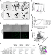
Figure 6. Mode of action of thrombin-derived C-terminal peptides.
(A) Electron microscopy analysis. P. aeruginosa and S. aureus bacteria was incubated for 2 h at 37°C with 30 µM of GKY25 and LL-37 and analyzed with electron microscopy. Scale bar represents 1 µm. Control; Buffer control. (B) Permeabilizing effects of peptides on E. coli. Bacteria were incubated with the indicated peptides at 30 µM and permeabilization was assessed using the impermeant probe FITC. (C) Helical content of the thrombin-derived C-terminal peptides GKY25 and VFR17 in presence of negatively charged liposomes (PA). The two peptides showed a marked helix induction upon addition of the liposomes. (D) CD spectra of GKY25 and VFR17 in Tris-buffer and in presence of LPS. For control, CD spectra for buffer and LPS alone are also presented. (E) Effects of the indicated peptides on liposome leakage. The membrane permeabilizing effect was recorded by measuring fluorescence release of carboxyfluorescein from PA (negatively charged) liposomes. The experiments were performed in 10 mM Tris-buffer, in absence and presence of 0.15 M NaCl. Values represents mean of triplicate samples. (F) Activities of corresponding C-terminal peptides of the indicated coagulation factors. Peptides were tested in RDA against the indicated bacteria. Bacteria (4×10⁶ cfu) were inoculated in 0.1% TSB agarose gels. Each 4 mm-diameter well was loaded with 6 µl of peptide at 100 µM. The zones of clearance correspond to the inhibitory effect of each peptide after incubation at 37°C for 18–24 h. (G) Overlay 3D-model showing the three coagulation factors thrombin, and factor X and IX. The C-terminal parts are indicated.

Similar articles

Cited by

References

    1. Lehrer RI, Ganz T. Cathelicidins: a family of endogenous antimicrobial peptides. Curr Opin Hematol. 2002;9:18–22. - PubMed
    1. Harder J, Glaser R, Schröder JM. Review: Human antimicrobial proteins effectors of innate immunity. J Endotoxin Res. 2007;13:317–338. - PubMed
    1. Zasloff M. Antimicrobial peptides of multicellular organisms. Nature. 2002;415:389–395. - PubMed
    1. Tossi A, Sandri L, Giangaspero A. Amphipathic, alpha-helical antimicrobial peptides. Biopolymers. 2000;55:4–30. - PubMed
    1. Yount NY, Bayer AS, Xiong YQ, Yeaman MR. Advances in antimicrobial peptide immunobiology. Biopolymers. 2006;84:435–458. - PubMed

Publication types

MeSH terms