. 2010 Apr 22;6(4):e1000860.
doi: 10.1371/journal.ppat.1000860.

Two coregulated efflux transporters modulate intracellular heme and protoporphyrin IX availability in Streptococcus agalactiae

Affiliations

Two coregulated efflux transporters modulate intracellular heme and protoporphyrin IX availability in Streptococcus agalactiae

Annabelle Fernandez et al. PLoS Pathog. .

Abstract

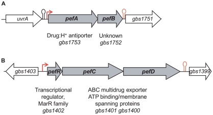
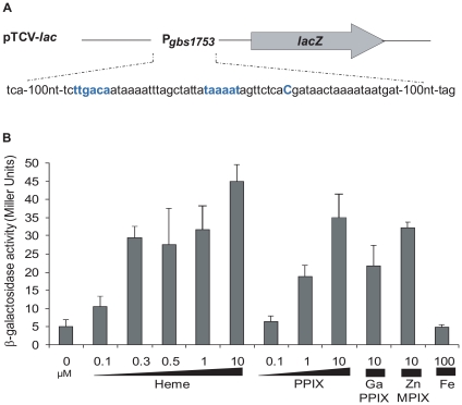
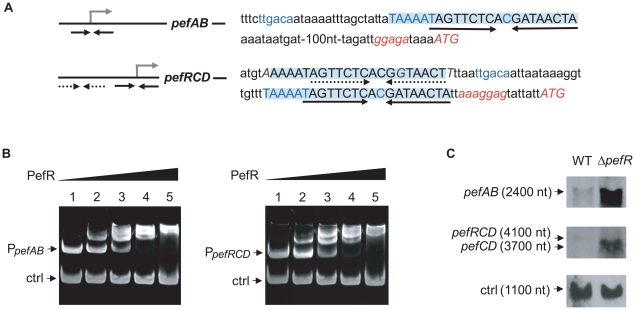
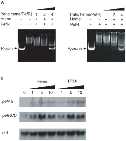
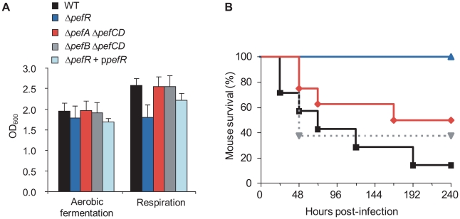
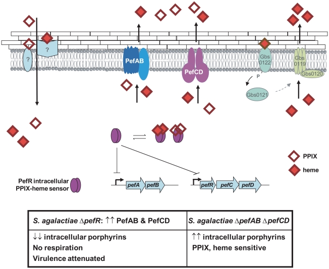
Streptococcus agalactiae is a major neonatal pathogen whose infectious route involves septicemia. This pathogen does not synthesize heme, but scavenges it from blood to activate a respiration metabolism, which increases bacterial cell density and is required for full virulence. Factors that regulate heme pools in S. agalactiae are unknown. Here we report that one main strategy of heme and protoporphyrin IX (PPIX) homeostasis in S. agalactiae is based on a regulated system of efflux using two newly characterized operons, gbs1753 gbs1752 (called pefA pefB), and gbs1402 gbs1401 gbs1400 (called pefR pefC pefD), where pef stands for 'porphyrin-regulated efflux'. In vitro and in vivo data show that PefR, a MarR-superfamily protein, is a repressor of both operons. Heme or PPIX both alleviate PefR-mediated repression. We show that bacteria inactivated for both Pef efflux systems display accrued sensitivity to these porphyrins, and give evidence that they accumulate intracellularly. The DeltapefR mutant, in which both pef operons are up-regulated, is defective for heme-dependent respiration, and attenuated for virulence. We conclude that this new efflux regulon controls intracellular heme and PPIX availability in S. agalactiae, and is needed for its capacity to undergo respiration metabolism, and to infect the host.


Conflict of interest statement

The authors have declared that no competing interests exist.

Figures

Figure 1
Figure 1. Genetic context of two loci for which expression is heme-induced.
A, gbs1753 gbs1752 (pefA pefB) and B, gbs1402 gbs1401 gbs1400 (pefR pefC pefD) loci. Bent arrows and lollipops indicate the mapped or putative promoters, and rho-independent terminators, respectively.
Figure 2
Figure 2. The pefAB locus is induced by porphyrin molecules: heme, PPIX, GaPPIX and ZnMPIX.
A. Schematic map of the transcriptional fusion PpefA-lacZ (same as Pgbs1753-lacZ). Sequence of the pefAB promoter region is displayed. The +1 transcriptional start and −10 and −35 motifs are in bold blue characters. B. Expression analysis of PpefA-lacZ by determination of β-galactosidase activity in early stationary phase. S. agalactiae was grown in BHI liquid medium in the presence of the indicated amount of candidate inducers. Strains harboring the pTCV-lac vector (promoterless negative control) did not show β-galactosidase activity during growth (0.4±0.1 Miller Units). Measurements were performed at least three times.
Figure 3
Figure 3. PefR is a repressor of pefAB and pefRCD loci.
A. A conserved 23-nucleotide motif is present once upstream of pefAB, and twice upstream of pefRCD and is highlighted. The IR present in each of the motifs is marked by arrows. The −10 and −35 motifs, and the mRNA pefAB and putative pefRCD start sites are in blue. Start codons and RBS sequence are in italics. One motif upstream of pefRCD (dotted arrows) differs from the other motifs by 3 nucleotides, which are in gray italics. B. PefR binds the pefAB and pefRCD promoter regions in gel shift assays. Two pmoles of pefAB or pefRCD promoter fragment were incubated in the presence of 0, 4, 17, 34, 63 pmoles of PefR (lanes 1 to 5) and 4.6 pmoles control fragment, corresponding to an 116-bp fragment of the pefA gene. C. Northern blot analyses of pefAB and pefRCD mRNA in the WT and pefR strains, using locus-specific probes (see Materials and Methods). As probe efficiencies and times of exposure differ for each target RNA, differences between pefAB and pefRCD levels are not comparable. ldhL mRNA (‘ctrl’) was used to control for RNA quantity.
Figure 4
Figure 4. PefR is a heme- and PPIX-modulated repressor of pefAB and pefRCD loci.
A. Gel mobility shift analysis of the effect of heme on PefR binding to pefAB (right) and pefRCD (left) promoter regions. 2 pmoles of PpefAB or PpefRCD fragments and 30 pmoles of PefR were mixed. Increased concentrations of heme were added in ratios indicated (keeping PefR constant). Assays were also performed with PPIX (Fig. S1A), giving similar results. On the two top panels, lanes 1–3 were juxtaposed to lanes 4–5, which were separated on the initial same gel. B. Northern blot analyses of pefAB and pefRCD expression in the presence of heme or PPIX in WT NEM316. Cultures were grown in the presence of 0, 1, 5 and 10 µM heme or PPIX, and harvested for total RNA extraction in early stationary phase. As probe efficiencies and times of exposure differ for each target RNA, differences between pefAB and pefRCD levels are not comparable. ldhL (gbs0947; ‘ctrl’) mRNA was used to control for RNA quantities.
Figure 5
Figure 5. The hrtAB locus is induced by higher heme concentrations than pefAB or pefRCD, and is not induced by PPIX.
A. Northern blot analyses of gbs0119 expression in the presence of heme or PPIX in WT NEM316. Cultures were grown in the presence of 0, 1, 5 and 10 µM heme or PPIX, and harvested for total RNA extraction in early stationary phase. ldhL (gbs0947; ‘crtl’) mRNA was used as RNA quantity control. The hybridization was performed on the same membrane as that used in Fig. 4B. B. Expression analysis of Pgbs0119-lacZ by determination of β-galactosidase activity in early stationary phase. S. agalactiae was grown in BHI liquid medium in the presence of different heme concentrations. Measurements were performed three or more times.
Figure 6
Figure 6. ΔpefAB and ΔpefCD mutants are affected in PPIX and heme sensitivity.
A. Growth curves of WT and mutant strains in the presence of heme. Cells were grown aerobically in M17G medium supplemented with 0 or 1 µM heme. WT (black circle), WT +1 µM heme (black square), ΔpefA ΔpefCD (red circle), ΔpefA ΔpefCD +1 µM heme (red square), ΔpefB ΔpefCD (gray circle, dashed line) and ΔpefB ΔpefCD +1 µM heme (gray square, dashed line). Results shown are representative of 3 experiments. Growth retardation of ΔpefA ΔpefCD or ΔpefB ΔpefCD was more pronounced with 2 and 4 µM heme. B. Photosensitivity of WT and mutant S. agalactiae strains grown in the presence of PPIX. Cells were grown until early stationary phase in M17G medium supplemented or not with 10 µM PPIX. Serial 10-fold dilutions (exponent is indicated) were exposed to 0, 10, or 50 minutes visible light. Plates were photographed after 24 h incubation.
Figure 7
Figure 7. PPIX intracellular accumulation in vivo.
A. Expression analysis of PpefA-lacZ by β-galactosidase activity determinations was performed in early stationary phase cells of WT and ΔpefA ΔpefCD strains. S. agalactiae and derivatives were grown in BHI liquid medium. Results represent the mean ± standard deviation from triplicate experiments. Asterisks denote statistically significant differences as determined by Student's t-test (p≤0.05). B. PPIX- or heme-dependent production of the E. faecalis catalase KatA in S. agalactiae WT, and ΔpefA ΔpefCD mutant strains. Cells were grown in M17G supplemented or not with 1 µM of PPIX or heme. The loading of equivalent amounts of protein was verified by Coomassie stained gels performed in parallel. KatA was detected in total S. agalactiae protein extracts by immunoblot assays. Results shown are representative of 3 experiments.
Figure 8
Figure 8. Physiological consequences of pef mutations.
A. Impact on respiration metabolism of deregulating pefAB pefCD expression by pefR inactivation. S. agalactiae WT, ΔpefA ΔpefCD, ΔpefB ΔpefCD, ΔpefR and ΔpefR transformed with vector containing a wild-type copy of pefRpefR + ppefR) strains were grown in respiration-permissive conditions (i.e., 1 µM heme and 10 µM menaquinone). S. agalactiae respiration growth is characterized by a gain in cell density . Absorbance (OD600) is shown on cultures after 22 h growth. Errors bars represent the standard deviation of three independent experiments. B. Impact on virulence of deregulating pefAB pefCD expression by pefR inactivation. Survival curves in adult mice infected with S. agalactiae WT (black square), ΔpefA ΔpefCD (red diamond), ΔpefB ΔpefCD (gray triangle, dashed line) or ΔpefR (blue triangle) strains. Differences in mortality between mice infected with the WT versus the ΔpefR mutant were statistically significant (p<0.001).
Figure 9
Figure 9. Model for pefAB and pefCD functions regulated by an intracellular sensor in S. agalactiae.
Extracellular heme and PPIX may be internalized by as yet unknown transporters. By homology with a recently described system in S. aureus , heme, but not PPIX, may bind to a putative external two-component receptor protein Gbs0122, the homolog of S. aureus HssS, resulting in gbs0119 gbs0120 (hrtAB) induction. Once inside the cell, free porphyrin molecules may encounter different binding proteins, including PefR. Apo-PefR binds to pefAB and pefRCD promoter regions to repress their expression. PefR-porphyrin binding releases PefR from DNA, to enable pefAB and pefRCD expression. The PefAB and PefCD loci mediate efflux of free heme and PPIX to avoid toxicity. Overexpression of PefAB and PefCD leads to heme depletion and respiration and virulence defects. Phenotypes of ΔpefR versus ΔpefAB ΔpefCD mutants are shown in the Table below.

Similar articles

Cited by

References

    1. Mense SM, Zhang L. Heme: a versatile signaling molecule controlling the activities of diverse regulators ranging from transcription factors to MAP kinases. Cell Res. 2006;16:681–692. - PubMed
    1. Shepherd M, Heath MD, Poole RK. NikA binds heme: a new role for an Escherichia coli periplasmic nickel-binding protein. Biochemistry. 2007;46:5030–5037. - PubMed
    1. Gilles-Gonzalez MA, Gonzalez G. Signal transduction by heme-containing PAS-domain proteins. J Appl Physiol. 2004;96:774–783. - PubMed
    1. Kumar S, Bandyopadhyay U. Free heme toxicity and its detoxification systems in human. Toxicol Lett. 2005;157:175–188. - PubMed
    1. Letoffe S, Heuck G, Delepelaire P, Lange N, Wandersman C. Bacteria capture iron from heme by keeping tetrapyrrol skeleton intact. Proc Natl Acad Sci U S A 2009 - PMC - PubMed

Publication types

MeSH terms