. 2010 Apr 22;6(4):e1000923.
doi: 10.1371/journal.pgen.1000923.

Cdh11 acts as a tumor suppressor in a murine retinoblastoma model by facilitating tumor cell death

Affiliations

Cdh11 acts as a tumor suppressor in a murine retinoblastoma model by facilitating tumor cell death

Mellone N Marchong et al. PLoS Genet. .

Abstract

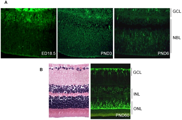
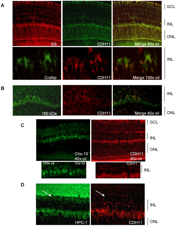
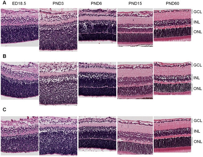
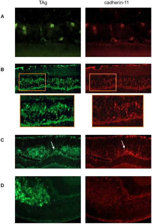
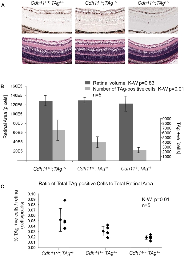
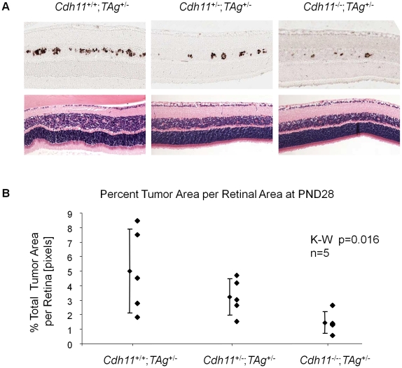
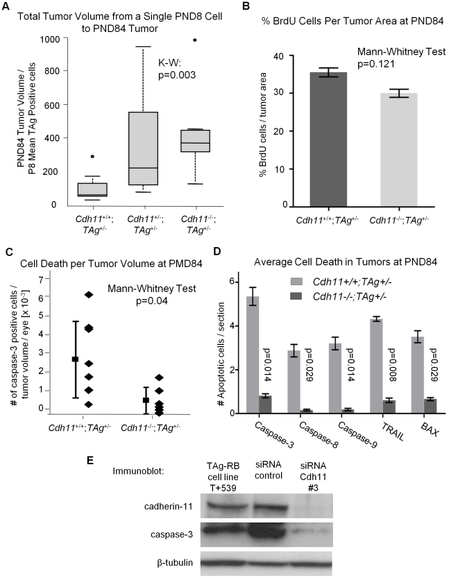
CDH11 gene copy number and expression are frequently lost in human retinoblastomas and in retinoblastomas arising in TAg-RB mice. To determine the effect of Cdh11 loss in tumorigenesis, we crossed Cdh11 null mice with TAg-RB mice. Loss of Cdh11 had no gross morphological effect on the developing retina of Cdh11 knockout mice, but led to larger retinal volumes in mice crossed with TAg-RB mice (p = 0.01). Mice null for Cdh11 presented with fewer TAg-positive cells at postnatal day 8 (PND8) (p = 0.01) and had fewer multifocal tumors at PND28 (p = 0.016), compared to mice with normal Cdh11 alleles. However, tumor growth was faster in Cdh11-null mice between PND8 and PND84 (p = 0.003). In tumors of Cdh11-null mice, cell death was decreased 5- to 10-fold (p<0.03 for all markers), while proliferation in vivo remained unaffected (p = 0.121). Activated caspase-3 was significantly decreased and beta-catenin expression increased in Cdh11 knockdown experiments in vitro. These data suggest that Cdh11 displays tumor suppressor properties in vivo and in vitro in murine retinoblastoma through promotion of cell death.


Conflict of interest statement

The authors have declared that no competing interests exist.

Figures

Figure 1
Figure 1. Expression of cadherin-11 in developing murine retina.
(A) Cadherin-11 was expressed in the differentiating layer at (embryonic day) ED18.5, by retinoblasts at (post natal day) PND3 and again in a differentiating layer at PND6. (B) In adult mice, (PND60) cadherin-11 expression was restricted to cell types of the INL, with high expression by Müller glia processes that span the entire retina. GCL: ganglion cell layer; INL: inner cell layer; ONL outer nuclear layer.
Figure 2
Figure 2. Co-expression of cadherin-11 and retinal cell types in adult retina.
(A, B) Cadherin-11 expression co-localizes with Müller glia cell bodies (CRALBP, 100× magnification), Müller glia cell processes (glutamine synthetase, 40× magnification) and horizontal cells (160 kDa, 40× magnification) (C, D) but not with bipolar (Chx-10, 40× and 100× magnification) or amacrine (HPC-1, 40× magnification) (white arrows) cells.
Figure 3
Figure 3. Retinal histology of Cdh11 +/+, Cdh11 +/-, and Cdh11 -/- littermates.
Hematoxylin and eosin (H&E) staining of 5 µm sections cut through the papillary-optic nerve plane. At developmental time points, ED18.5, PND3, PND6, PND15 (data not shown) and PND60 (adult), no gross retinal phenotypic differences were observed between (A) Cdh11 +/+, (B) Cdh11 +/-, and (C) Cdh11 -/- littermates.
Figure 4
Figure 4. Gradual loss of cadherin-11 expression in TAg-RB tumors.
(A) At PND9 TAg-RB mice displayed single TAg-positive cells (green) also positive for cadherin-11 (red). (B) At PND28 TAg-RB mice displayed multifocal tumors (clusters) which stained positive for TAg (green). Some of these multifocal tumors lost cadherin-11 expression (left cluster in box), while some retained expression (right cluster in box), suggesting a partial loss of cadherin-11 expression from PND28 tumors. (C) At PND35, regions of tumors that were positive for TAg were completely negative for cadherin-11 and adjacent normal cells retained cadherin-11 expression (arrow). (D) By PND140, entire tumors showed no cadherin-11 expression.
Figure 5
Figure 5. Cdh11 genomic copy number correlates with number of TAg-positive cells (origin of tumors in TAg-RB mice) at PND8.
(A) Representative sections of Cdh11 +/+;TAg +/-, Cdh11 +/-;TAg +/-, and Cdh11- /-;TAg +/- genotypes by H&E stain and TAg staining. The single TAg-positive cells in the INL of the retina are reduced in number with reduced Cdh11 allele dosage. H&E staining reveals no major phenotypic differences between the three genotypes. (B) Manual counts of TAg-positive cells per retinal area were extrapolated to the entire retina. The total number of TAg-positive cells of origin of retinoblastoma was 2-fold and 3-fold less (p = 0.01) (light grey bars) when one or two alleles of Cdh11 were respectively lost, as compared to mice with normal Cdh11. The retinal size (dark grey bars) was similar (p = 0.83) between the Cdh11 genotypes. (C) The ratio of TAg-positive cells to total retinal area was significantly reduced with reduced Cdh11 gene dose (p = 0.01). The Kruskal-Wallis Test was used to assess difference between groups and error bars represent standard deviations.
Figure 6
Figure 6. At PND28, fewer multifocal tumors developed when Cdh11 alleles were lost.
(A) A distinct Cdh11 loss phenotype was observed from representative sections of TAg and H&E stains. Fewer TAg-positive multifocal tumors were present in mice with mutant Cdh11 alleles; H&E showed more advanced tumors in Cdh11 +/+ mice than in Cdh11 +/- or Cdh11- /- mice. (B) The number of multifocal tumors was significantly less (p = 0.016) in mice with Cdh11 allelic loss, correlating with fewer tumor initiating cells at PND8. Total tumor volume was calculated using image J software measuring tumor area (TAg stained region) as a percentage of retinal area (manually traced) for every 60th section (approximately 300 µm apart) through the eye and extrapolated to the entire retina.
Figure 7
Figure 7. At PND84, total tumor volume was similar in all three genotypes.
(A) Representative H&E and TAg stained sections showed large tumors originating from the INL of the retina. Tumors were composed of disorganized cells, rosette formations and disrupted laminated layers. No gross phenotypic differences were observed in different genotypes on H&E stained sections. (B) Retinal area and tumor area of every 60th section were tabulated and extrapolated to the entire retina. Total tumor volume per genotype was not statistically different (p = 0.26), but total retinal areas were significantly larger when Cdh11 was lost (p = 0.01). (C) To accommodate for varying retinal size per genotype, total tumor volume was represented as a percentage total retinal area in all mice, showing no statistical difference between genotypes (p = 0.07), although a strong trend is observed, perhaps due to overall larger retinas. This suggested faster growing tumors in mice with Cdh11 loss, since there were fewer tumor-originating cells and consequently fewer multifocal tumors initially (PND28). Tumor volume was calculated as described in Figure 5.
Figure 8
Figure 8. Allelic loss of Cdh11 led to faster growing tumors due to decreased cell death.
(A) The number of single tumor initiating cells at PND8 was estimated by averaging the total number of TAg-positive cells in 5 mice per genotype. The ratio of tumor volume in pixels at PND84 to the average number of TAg-positive cells per eye at PND8 for the three genotypes was used to estimate tumor growth rate. A significant difference in growth rate between groups was observed (p = 0.003), with a 3-fold increase between Cdh11 -/- and wild type mice. After controlling for larger retinas (Figure 6B), growth rate remained significantly larger in Cdh11 -/- mice (data not shown). (B) Cdh11+/+;TAg+/- and Cdh11-/-;TAg+/- PND84 eyes were stained for BrdU incorporation (n = 6 per group). Proliferation was analyzed by averaging slides for the % BrdU positive cells (in pixels) per tumor area (determined by TAg staining in pixels). No significant difference was observed (p = 0.121). (C) Every 60th section of Cdh11+/+;TAg+/- (n = 8) and Cdh11-/-;TAg+/- (n = 6) PND84 eyes, was counted for tumor cells positive for activated caspase-3, extrapolated to the entire retina and represented as a ratio to tumor volume per eye. The number of dying cells in tumors of mice with normal Cdh11 alleles are significantly more abundant (p = 0.04) than in tumors of mice with mutant Cdh11 alleles. (D) To further support this data, we analyzed for the expression of five pro-apoptotic proteins (activated caspase-3, 8, 9, Trail and Bax) in an additional cohort of Cdh11+/+;TAg+/- and Cdh11-/-;TAg+/- PND84 animals. Staining and analysis were performed as performed previously. Results revealed between five to ten times less apoptotic activity in TAg-RB tumors null for Cdh11 (p = 0.014, 0.029, 0.014, 0.008, and 0.029, respectively; n = 4 per group). (E) Cadherin-11 was knocked down using stealth siRNA in a cadherin-11 expressing cell line derived from TAg-RB tumors, T +539. Knockdown of Cdh11 clearly decreased caspase-3 expression compared to control.

Similar articles

Cited by

References

    1. Gallie BL, Campbell C, Devlin H, Duckett A, Squire JA. Developmental basis of retinal-specific induction of cancer by RB mutation. Cancer Res. 1999;59:1731s–1735s. - PubMed
    1. Dimaras H, Coburn B, Pajovic S, Gallie BL. Loss of p75 neurotrophin receptor expression accompanies malignant progression to human and murine retinoblastoma. Mol Carcinog. 2006;45:333–343. - PubMed
    1. Dimaras H, Khetan V, Halliday W, Orlic M, Prigoda NL, et al. Loss of RB1 induces non-proliferative retinoma; increasing genomic instability correlates with progression to retinoblastoma. Hum Mol Genet. 2008. - PubMed
    1. Squire J, Gallie BL, Phillips RA. A detailed analysis of chromosomal changes in heritable and non-heritable retinoblastoma. Hum Genet. 1985;70:291–301. - PubMed
    1. Chen D, Gallie BL, Squire JA. Minimal regions of chromosomal imbalance in retinoblastoma detected by comparative genomic hybridization. Cancer Genet Cytogenet. 2001;129:57–63. - PubMed

Publication types