

PAX3 expression in normal skin melanocytes and melanocytic lesions (naevi and melanomas)
- PMID: 20421967
- PMCID: PMC2858648
- DOI: 10.1371/journal.pone.0009977
PAX3 expression in normal skin melanocytes and melanocytic lesions (naevi and melanomas)
Abstract
Background: Cutaneous Malignant Melanoma is an aggressive form of skin cancer, arising in cutaneous melanocytes. The transcription factor PAX3 regulates melanocyte specification from neural crest cells during development but expression in differentiated melanocytes is uncertain. By contrast it is frequently found in melanomas and naevi and is a marker for melanoma staging and detection. In this study we analysed the expression of PAX3 across the spectrum of melanocytic cells, from normal melanocytes to cells of benign and malignant lesions to better assess its function in these various tissues. Pax3 and PAX3 (italicized) refer to the mouse and human gene, respectively; whereas Pax3 and PAX3 (non-italicized) refer to the corresponding mouse and human protein.
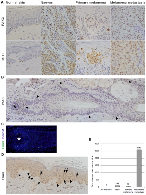
Methodology and principal findings: PAX3 expression was analysed by immunohistochemistry and qRT-PCR. Immunofluorescence was used for co-expression with differentiation, migration and survival markers. As expected PAX3 expression was observed in naevi and melanoma cells. It was also found in melanocytes of normal skin where it co-expressed with melanocyte markers, MITF and MLANA. Co-expression with its downstream target, antiapoptotic factor BCL2L1 confirms PAX3 as a cell survival regulator. PAX3 was also co-expressed with melanoma cell migration marker MCAM in dermal naevi and melanoma cell nests, but this downstream target of PAX3 was not present in normal epidermal melanocytes, suggesting differential roles for PAX3 in normal epidermal melanocytes and melanoma cells. Most interestingly, a proportion of PAX3-positive epidermal melanocytes in normal skin show HES1 and Ki67 co-expression, indicating their less differentiated proliferative phenotype.
Conclusions and significance: Our results suggest that a previously identified role for PAX3, that of regulator of an undifferentiated plastic state, may operate in melanocytes of normal skin. This role, possibly required for cellular response to environmental stimuli, may contribute to formation and development of melanocytic lesions in which PAX3 expression is prominent.
Conflict of interest statement
Figures


Similar articles
-
Differential PAX3 functions in normal skin melanocytes and melanoma cells.Biochem Biophys Res Commun. 2011 Aug 12;411(4):832-7. doi: 10.1016/j.bbrc.2011.07.053. Epub 2011 Jul 23. Biochem Biophys Res Commun. 2011. PMID: 21802410
-
Expression of genes for microphthalmia isoforms, Pax3 and MSG1, in human melanomas.Cell Mol Biol (Noisy-le-grand). 1999 Nov;45(7):1075-82. Cell Mol Biol (Noisy-le-grand). 1999. PMID: 10644012
-
PAX3 expression in primary melanomas and nevi.Mod Pathol. 2008 May;21(5):525-30. doi: 10.1038/modpathol.3801019. Epub 2008 Mar 7. Mod Pathol. 2008. PMID: 18327212 Free PMC article.
-
PAX3 across the spectrum: from melanoblast to melanoma.Crit Rev Biochem Mol Biol. 2009 Jun;44(2-3):85-97. doi: 10.1080/10409230902755056. Crit Rev Biochem Mol Biol. 2009. PMID: 19401874 Review.
-
Pigmentation PAX-ways: the role of Pax3 in melanogenesis, melanocyte stem cell maintenance, and disease.Pigment Cell Melanoma Res. 2008 Dec;21(6):627-45. doi: 10.1111/j.1755-148X.2008.00514.x. Pigment Cell Melanoma Res. 2008. PMID: 18983540 Free PMC article. Review.
Cited by
-
Defining the transcriptome of PIK3CA-altered cells in a human capillary malformation using single cell long-read sequencing.Sci Rep. 2024 Oct 25;14(1):25440. doi: 10.1038/s41598-024-72167-8. Sci Rep. 2024. PMID: 39455600 Free PMC article.
-
Markers of circulating tumour cells in the peripheral blood of patients with melanoma correlate with disease recurrence and progression.Br J Dermatol. 2013 Jan;168(1):85-92. doi: 10.1111/bjd.12057. Epub 2012 Nov 15. Br J Dermatol. 2013. PMID: 23013138 Free PMC article.
-
Cell cycle regulatory markers in melanoma: New strategies in diagnosis and treatment.Med J Islam Repub Iran. 2019 Sep 14;33:96. doi: 10.34171/mjiri.33.96. eCollection 2019. Med J Islam Repub Iran. 2019. PMID: 31696090 Free PMC article. Review.
-
The role of microRNAs and long non-coding RNAs in the pathology, diagnosis, and management of melanoma.Arch Biochem Biophys. 2014 Dec 1;563:60-70. doi: 10.1016/j.abb.2014.07.022. Epub 2014 Jul 24. Arch Biochem Biophys. 2014. PMID: 25065585 Free PMC article. Review.
-
Exosomes Derived from Hypertrophic Scar Fibroblasts Suppress Melanogenesis in Normal Human Epidermal Melanocytes.Int J Mol Sci. 2024 Jun 30;25(13):7236. doi: 10.3390/ijms25137236. Int J Mol Sci. 2024. PMID: 39000342 Free PMC article.
References
-
- Barber TD, Barber MC, Cloutier TE, Friedman TB. PAX3 gene structure, alternative splicing and evolution. Gene. 1999;237:311–319. - PubMed
-
- Barr FG, Fitzgerald JC, Ginsberg JP, Vanella ML, Davis RJ, et al. Predominant expression of alternative PAX3 and PAX7 forms in myogenic and neural tumor cell lines. Cancer Res. 1999;59:5443–5448. - PubMed
-
- Galibert MD, Yavuzer U, Dexter TJ, Goding CR. Pax3 and regulation of the melanocyte-specific tyrosinase-related protein-1 promoter. J Biol Chem. 1999;274:26894–26900. - PubMed
-
- Muratovska A, Zhou C, He S, Goodyer P, Eccles MR. Paired-Box genes are frequently expressed in cancer and often required for cancer cell survival. Oncogene. 2003;22:7989–7997. - PubMed
Publication types
MeSH terms
Substances
LinkOut - more resources
Full Text Sources
Medical
Research Materials
