. 2010 Apr 20;5(4):e10248.
doi: 10.1371/journal.pone.0010248.

Drosophila SAF-B links the nuclear matrix, chromosomes, and transcriptional activity

Affiliations

Drosophila SAF-B links the nuclear matrix, chromosomes, and transcriptional activity

Catalina Alfonso-Parra et al. PLoS One. .

Abstract

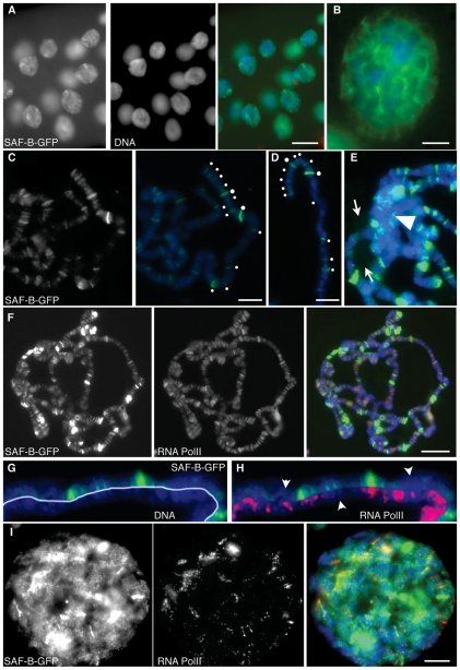
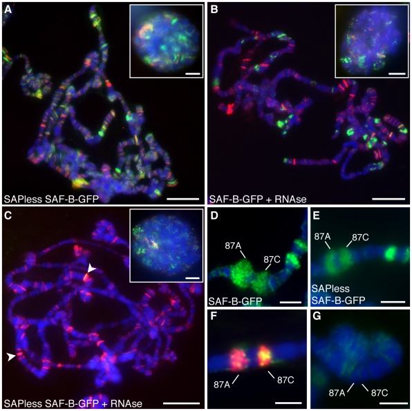
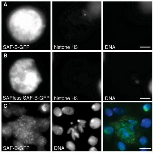
Induction of gene expression is correlated with alterations in nuclear organization, including proximity to other active genes, to the nuclear cortex, and to cytologically distinct domains of the nucleus. Chromosomes are tethered to the insoluble nuclear scaffold/matrix through interaction with Scaffold/Matrix Attachment Region (SAR/MAR) binding proteins. Identification and characterization of proteins involved in establishing or maintaining chromosome-scaffold interactions is necessary to understand how the nucleus is organized and how dynamic changes in attachment are correlated with alterations in gene expression. We identified and characterized one such scaffold attachment factor, a Drosophila homolog of mammalian SAF-B. The large nuclei and chromosomes of Drosophila have allowed us to show that SAF-B inhabits distinct subnuclear compartments, forms weblike continua in nuclei of salivary glands, and interacts with discrete chromosomal loci in interphase nuclei. These interactions appear mediated either by DNA-protein interactions, or through RNA-protein interactions that can be altered during changes in gene expression programs. Extraction of soluble nuclear proteins and DNA leaves SAF-B intact, showing that this scaffold/matrix-attachment protein is a durable component of the nuclear matrix. Together, we have shown that SAF-B links the nuclear scaffold, chromosomes, and transcriptional activity.


Conflict of interest statement

Competing Interests: The authors have declared that no competing interests exist.

Figures

Figure 1
Figure 1. Structure of the gene and gene products of Drosophila SAF-B.
(A) Human SAF-B1 and SAF-B2 possess the same domains as the Drosophila homologue, CG6995. Characterized domains (SAP, RRM) are shown, as well as regions of notable low sequence complexity (K-, G-, R-, and E-rich). (B) NetPhos 2.0 algorithm identification of potential phosphorylation sites, height of bar indicates probability of phosphorylation. Phosphopeptides confirmed in PhosphoPep database are in black, along with putative responsible kinase based on consensus match (asterisks indicate no clear consensus match); gray bar indicates Doa consensus without supporting PhosphoPep support. Three structural determination algorithms (gray bars are PONDR VL-XT, DisEMBL, and IUPred) show extensive predicted intrinsically disordered domains. Averaging scores (black line) shows the only predicted ordered domains are the SAP and RRM domains. (C) Gray bar at top represents genomic DNA, and locations of oligonucleotide primers described in Materials and Methods are shown (1-6). Identified mRNA species (shown as alternating thick exonic and thin intronic gray bars) encode two different protein products (conceptual translation products are shown as thick black bars with colored domains as in (a)). Isoform B is annotated at Flybase, as are two other forms for which we could find no supportive data - isoform A includes intron 5 (asterisk), and isoform C includes introns 5 and 6 (double-asterisk) and is missing exons 1-3 and introns 1-3. Our analyses also identified a novel form, D, which does not possess the RRM, one of the G-rich domains, and contains a shorter R/E-rich domain.
Figure 2
Figure 2. Expression profile of saf-b.
(A) Reverse-Transcriptase Polymerase Chain Reaction shows expression in all life stages, and in soma (heads) and mixed soma/germ (bodies). Primers 2 and 5 were used for the B form, and 2 and 6 were used for the D form. 18S rRNA was used as extraction, reverse transcriptase, PCR, and loading control. Lanes 1–7 are embryos, larvae, pupae, female heads, female decapitated bodies, male heads, and male decapitated bodies. (B–H) embryonic stages showing expression in precellularized syncytial embryos (B), mid-cellularized blastoderm embryos (C), gastrula (D), germ-band elongated (E) and retracted (F) embryos, and late-stage embryos with noticeably intense staining in the central (G) and peripheral (H) nervous system. (I-K) Expression in third-instar larval brains (I), leg imaginal discs (J), and eye-antennal discs (K). (L-M) Expression in the germline of males (L) and females (M). Expression is not detected in the germline stem cells (upper-rightmost tips in both testes and germaria), but is evident in developing spermatocytes, and nurse cells and oocytes.
Figure 3
Figure 3. Localization of SAF-B fusion proteins in cells.
(A) Carboxy-terminal GFP protein fusion to SAF-B (SAF-B-GFP) in an S2 cell nucleus. Independent channels for immunodetection of SAF-B-GFP and DAPI fluorescence of DNA, and the merge. General nucleoplasmic staining is evident, as well as more intense focal accumulations of protein. SAF-B is not enriched or excluded from the DAPI-bright heterochromatic compartment, but it is clearly excluded from the DAPI-dim nucleolus. (B) Immunodetection of Nuclear Pore Complex proteins p110 and p95, an integral membrane nuclear pore complex, fusion with images from (A), and an image with increased magnification showing no overlap between nuclear pore complexes and SAF-B foci. (C) Immunodetection of SAF-B-GFP and DAPI stained DNA, and the merge. Three S2 cells with different levels of SAF-B-GFP expression all show same nucleoplasmic and focal localization. (D) Amino-terminal GFP protein fusion to SAF-B (GFP-SAF-B) in an S2 cell nucleus. Independent channels for immunodetection of GFP-SAF-B, DAPI fluorescence of DNA, and the merge. Distribution of GFP-SAF-B is identical to that of the carboxy-terminal fusion shown in (A). (E) Distribution of SAF-B-GFP in early pre-determined embryonic nuclei, showing general nucleoplasmic and focal localization, as in S2 cells. (F) Distribution of SAF-B-GFP in larval neuroblast nuclei, showing general nucleoplasmic and focal localization, as in S2 cells and early embryos. Scale bar 2 µm (A, B, D, F) or 5 µm (C, E).
Figure 4
Figure 4. Distribution of SAF-B missing the conserved DNA-binding SAP domain.
(A) Truncated SAF-B protein, lacking the DNA-binding SAP domain, fused to GFP, in larval neuroblast nuclei. Independent channels are shown for immunodetection of SAF-B-GFP, DAPI fluorescence of DNA, and the merge. Few smaller foci (as seen in Fig. 3) are often replaced by one or more larger foci. These foci are often connected via continual “threadlike” structures of staining. (B-D) as in (A), SAPless SAF-B-GFP channel only. Scale bar 2 µm.
Figure 5
Figure 5. Distribution of SAF-B-GFP in polytene larval salivary gland nuclei.
(A) Immunodetection of SAF-B-GFP, DAPI staining of DNA, and the merge. The same nucleoplasmic and focal distribution seen in diploid cells are apparent. Additionally, more intense foci and threadlike continua are seen to connect foci. (B) Increased magnification to show continua. Brighter foci are at confluences of continua. (C) Squashed nuclei showing distribution of SAF-B-GFP, and merge with DAPI-stained DNA. SAF-B associates with specific bands on polytene chromosomes. White dots highlight SAF-B bands at the tip of the X chromosome. (D) A different polytene X chromosome, labelled as in (C), showing consistency of banding pattern. (E) The chromocenter (arrowhead) of salivary gland chromosomes does not show enhanced or reduced localization of SAF-B-GFP, although some foci are seen, and the nucleolus (arrows) shows only very low level of SAF-B-GFP. (F) Immunodetection of SAF-B-GFP, RNA Polymerase II (Ser2-PO4), and merge with DAPI-stained DNA. Extensive areas of overlap of both epitopes are evident, as are bands with detection of only SAF-B-GFP or RNA Polymerase II. (G) Immunodetection of SAF-B-GFP counterstained with DAPI to reveal salivary gland chromosome bands. SAF-B localized primarily to interband regions. (H) Immunodetection of SAF-B-GFP and DAPI staining of DNA as in (G), with immunodetection of RNA Polymerase II (Ser2-PO4), showing most bands of SAF-B overlap with RNA Polymerase II, but some bands of only one detectable epitope are apparent (arrowheads). (I) Immunodetection of SAF-B-GFP and RNA Polymerase II (Ser2-PO4) in whole-mount salivary gland nuclei, and merge with DAPI-stained DNA. Scale bar 50 µm (A), 10 µm (B–E, I), or 20 µm (F).
Figure 6
Figure 6. RNA- and transcription-dependent localization of SAF-B.
(A) Immunodetection of SAF-B-GFP lacking the DNA-binding SAP domain (green) and RNA Polymerase II (Ser2-PO4) (red) merged with DAPI-staining of DNA (blue). (B) Immunodetection of full-length SAF-B-GFP (green) and RNA Polymerase II (Ser2-PO4) (red) merged with DAPI-staining of DNA (blue) after treatment with RNAseA. (C) Immunodetection of SAF-B-GFP lacking the DNA-binding SAP domain (green) and RNA Polymerase II (Ser2-PO4) (red) merged with DAPI-staining of DNA (blue) after treatment with RNAseA. Arrowheads point to bands of retained SAF-B. Insets in A-C are whole mount nuclei. (D) Immunodetection of SAF-B-GFP at cytological bands 87A-C (green) merged with DAPI-stained DNA (blue), after 15 minute heat shock to induce expression. (E) Immunodetection of SAF-B-GFP lacking the DNA-binding SAP domain merged with DAPI-stained DNA after 15 minute heat shock, showing recruitment of SAPless SAF-B-GFP to the newly transcribed DNA. (F) Immunodetection of SAF-B-GFP (green) at cytological bands 87A-C (green) is reduced after RNAse treatment, although RNA Polymerase II (Ser2-PO4) (red) is still present. (G) Immunodetection of SAF-B (green) is negligible prior to heat shock induction of transcription. Scale bar 20 µm or 10 µm (inset images).
Figure 7
Figure 7. Nuclear extraction reveal SAF-B is a durable component of the nuclear matrix.
(A) Immunodetection of SAF-F-GFP and histone H3, and DAPI-staining of DNA after salt, detergent, and DNAseI extraction of mildly fixed nuclei. DNA and histone (a soluble nuclear and chromosome-bound component) are both removed entirely, while the SAF-B is retained. Scale bar 2 µm. (B) SAF-B truncated protein, lacking the SAP DNA-binding domain, is also retained in the nuclear matrix after extraction. Scale bar 2 µm. (C) Immunodetection of SAF-B in diploid mitotic neuroblast cells, DAPI-stained DNA, and merge. SAF-B is found in foci throughout the cytoplasm, and is not detectably associated with chromosomes. Scale bar 2 µm (A, B) or 5 µm (C).

Similar articles

Cited by

References

    1. Sutherland H, Bickmore WA. Transcription factories: gene expression in unions? Nat Rev Genet. 2009;10:457–466. - PubMed
    1. Solovei I, Cavallo A, Schermelleh L, Jaunin F, Scasselati C, et al. Spatial preservation of nuclear chromatin architecture during three-dimensional fluorescence in situ hybridization (3D-FISH). Exp Cell Res. 2002;276:10–23. - PubMed
    1. Berger AB, Cabal GG, Fabre E, Duong T, Buc H, et al. High-resolution statistical mapping reveals gene territories in live yeast. Nat Methods. 2008;5:1031–1037. - PubMed
    1. Clegg NJ, Honda BM, Whitehead IP, Grigliatti TA, Wakimoto B, et al. Suppressors of position-effect variegation in Drosophila melanogaster affect expression of the heterochromatic gene light in the absence of a chromosome rearrangement. Genome. 1998;41:495–503. - PubMed
    1. Hessler AY. V-Type Position Effects at the Light Locus in Drosophila Melanogaster. Genetics. 1958;43:395–403. - PMC - PubMed

Publication types