. 2010 Apr 20;5(4):e10260.
doi: 10.1371/journal.pone.0010260.

Phosphorylation of AATYK1 by Cdk5 suppresses its tyrosine phosphorylation

Affiliations

Phosphorylation of AATYK1 by Cdk5 suppresses its tyrosine phosphorylation

Koji Tsutsumi et al. PLoS One. .

Abstract

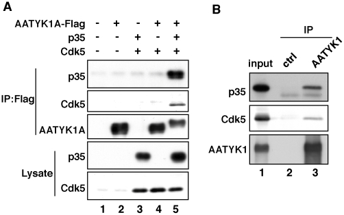
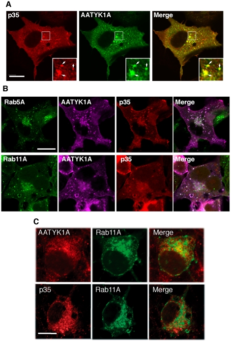
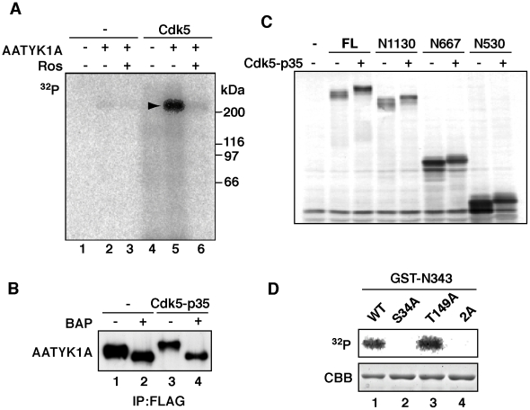
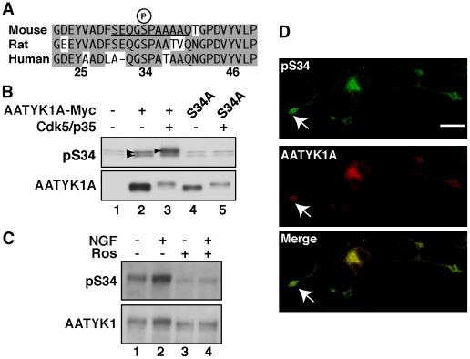
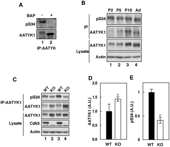
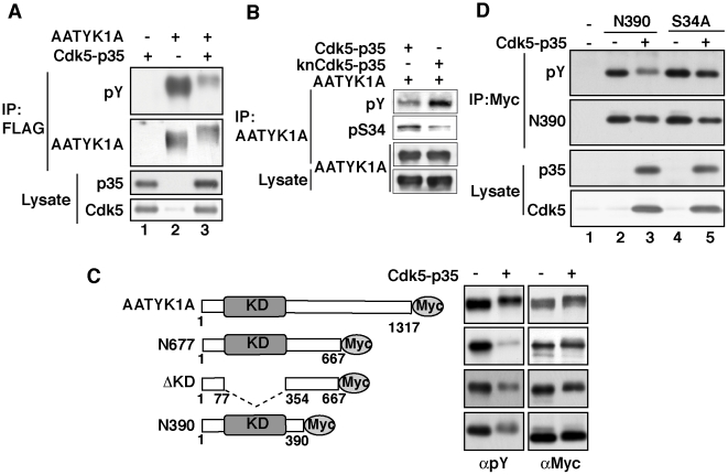
Apoptosis-associated tyrosine kinase 1 (AATYK1), a novel serine/threonine kinase that is highly expressed in the brain, is involved in neurite extension and apoptosis of cerebellar granule neurons; however, its precise function remains unknown. In this study, we investigated the interaction of AATYK1A with Cyclin-dependent kinase 5 (Cdk5)/p35, a proline-directed protein kinase that is predominantly expressed in neurons. AATYK1A bound to the p35 activation subunit of Cdk5 in cultured cells and in mouse brains and colocalized with p35 on endosomes in COS-7 cells. AATYK1A was phosphorylated at Ser34 by Cdk5/p35 in vitro, in cultured neurons and in mouse brain. In PC12D cells, Ser34 phosphorylation increased after treatment with nerve growth factor and phosphorylated AATYK1A accumulated in growth cones of PC12D cells. Ser34 phosphorylation suppressed the tyrosine phosphorylation of AATYK1A by Src family kinases. These results suggest a possibility that AATYK1A plays a role in early to recycling endosomes and its function is regulated by phosphorylation with Cdk5 or Src-family kinases.


Conflict of interest statement

Competing Interests: The authors have declared that no competing interests exist.

Figures

Figure 1
Figure 1. Binding of AATYK1A to Cdk5/p35.
(A) Coimmunoprecipitation of Cdk5/p35 with AATYK1A in HEK293 cells. AATYK1A-Flag was coexpressed with p35 and/or Cdk5 in HEK293 cells and immunoprecipitated from cell lysates using the anti-Flag antibody. p35 and Cdk5 were detected in the anti-Flag immunoprecipitates by immunoblotting using anti-p35 and anti-Cdk5 antibodies. (B) Coimmunoprecipitation of Cdk5/p35 from mouse brain extracts using the anti-AATYK1 antibody. AATYK1 was immunoprecipitated from a mouse brain extract (10 weeks). p35 and Cdk5 were detected in the AATYK1 immunoprecipitates using the anti-p35 and anti-Cdk5 antibodies.
Figure 2
Figure 2. Colocalization of p35 with AATYK1A in early and recycling endosomes.
(A) Colocalization of AATYK1A and p35 in COS-7 cells. COS-7 cells were transfected with AATYK1A-Myc together with p35 and Cdk5. AATYK1A and p35 were detected by immunostaining with the anti-Myc antibody and anti-p35 antibody, followed by incubation with Alexa 488-conjugated anti-mouse IgG and Alexa 548-conjugated anti-rabbit antibody, respectively. A merged image is shown in the right panel. Insets represent higher magnifications and arrows indicate the colocalization. Scale bar, 20 µm. (B) Localization of AATYK1A and p35 in early and recycling endosomes. COS-7 cells were transfected with AATYK1A-Myc, p35, Cdk5, and either EGFP-Rab5A (as an early-endosome marker) or EGFP–Rab11A (as a recycling-endosome marker). After 24 h of transfection, cells were fixed and stained with anti-Myc and anti-p35 antibodies, as described above, and were observed using a confocal microscope. Scale bar, 10 µm. (C) Localization of AATYK1 and p35 in endosomes in cultured cortical neurons. Rat brain cortical neurons at DIV5 were transfected with EGFP-Rab11A (middle panels). The cells were immunostained with anti-AATYK1 and p35 (C19) 24 h after transfection, followed by Alexa 546-conjugated anti-rabbit secondary antibody (left panels). Merge is shown in right panels. Bar, 10 µm.
Figure 3
Figure 3. Phosphorylation of AATYK1A at Ser34 by Cdk5.
(A) In vitro phosphorylation of AATYK1A by Cdk5/p35. AATYK1A-Flag was expressed in HEK293 cells and isolated by immunoprecipitation using the anti-Flag antibody. AATYK1A-Flag was incubated with Cdk5/p35 and [γ-32P]ATP in the presence or absence of 50 µM roscovitine (Ros) at 35°C for 30 min. The arrowhead indicates the phosphorylation of AATYK1A. (B) Dephosphorylation of AATYK1A by treatment with alkaline phosphatase. AATYK1A-Flag expressed in the presence (+) or absence (–) of Cdk5/p35 in HEK293 cells was isolated by immunoprecipitation using the anti-Flag antibody. Immunoprecipitates were treated with bacterial alkaline phosphatase (BAP) at 37°C for 30 min and immunoblotted with the anti-Flag antibody. (C) Phosphorylation of amino-terminal fragments of AATYK1A by Cdk5. The full-length (FL) and amino-terminal fragments of AATYK1A-Myc, N1130, N667, and N530, were expressed in HEK293 cells in the presence (+) or absence (–) of Cdk5/p35. HEK293 cell extracts were immunoblotted using the anti-Myc antibody. (D) Ser34 is a Cdk5 phosphorylation site of AATYK1A. The GST-N343 fragment or its Ala mutant at Ser34 (S34A), Thr149 (T149A), or both (2A) was incubated with purified Cdk5/p35 in the presence of [γ-32P]ATP at 35°C for 30 min. Phosphorylation was detected using autoradiography after SDS–PAGE. Coomassie Brilliant Blue (CBB) staining of GST-N343 is shown in the lower panel.
Figure 4
Figure 4. Generation of the anti-pSer34-specific antibody and Ser34 phosphorylation of AATYK1 in PC12D cells.
(A) Amino-acid sequences of mouse, rat, and human AATYK1A around Ser34. A synthetic peptide corresponding to the mouse AATYK1A amino-acid residues 29–39 (the Ser34 phosphorylation site is underlined) was used for rabbit immunization. (B) Specificity of the anti-pS34 antibody. COS-7 cells were transfected with AATYK1A or its S34A mutant in the presence (+) or absence (–) of Cdk5/p35. Cell extracts were immunoblotted with the anti-pS34 antibody or anti-Myc antibody for AATYK1A. (C) Phosphorylation of AATYK1 at Ser34 in PC12D cells. PC12D cells were treated with 50 ng/ml NGF for 24 h in the presence or absence of 20 µM roscovitine (Ros). AATYK1 was immunoprecipitated in PC12D cells using the anti-AATYK1 antibody and was subjected to immunoblotting with the anti-pS34 or anti-AATYK1 antibodies. (D) Immunofluorescent staining of PC12D cells using the anti-pS34 antibody. PC12D cells expressing AATYK1A-Myc were treated with NGF for 24 h and double labeled with the anti-pS34 (top panel) and anti-Myc (AATYK1A, middle panel) antibodies. A merged image is shown in the lower panel. The growth cone is indicated by an arrow. Scale bar, 20 µm.
Figure 5
Figure 5. Cdk5-mediated in vivo Ser34 phosphorylation.
(A) Phosphorylation of AATYK1 in mouse brain. AATYK1 was immunoprecipitated from mouse brain extracts and incubated in the presence (+) or absence (–) of bacterial alkaline phosphatase (BAP). The anti-pS34 reaction is shown in the upper panel and the anti-AATYK1 reaction is shown in the lower panel. (B) Phosphorylation of AATYK1 in mouse brain during early postnatal development. AATYK1 was immunoprecipitated from mouse brain extracts at P2, P5, P10, and six weeks of age (Ad) using the anti-AATYK1 antibody. The immunoprecipitates were immunoblotted using the anti-pS34 (top panel) and anti-AATYK1 (second panel) antibodies. Immunoblots of brain extracts using anti-AATYK1 and anti-actin antibodies are also shown in the lower panels. (C) Phosphorylation of AATYK1 at Ser34 in Cdk5–/– mouse brain. AATYK1 was immunoprecipitated from brain extracts of Cdk5–/– mice at embryonic day 18.5 (E18.5) and immunoblotted using the anti-pS34 antibody (top panel). Immunoblots of the brain extracts using anti-AATYK1 (third panel), anti-Cdk5 (fourth panel), and anti-actin (bottom panel) antibodies. Quantification of AATYK1 and pS34 is shown in (D) and (E), respectively. Bars indicate the means ± S.E. of three independent experiments (n = 3; * P<0.05; t test).
Figure 6
Figure 6. Phosphorylation of AATYK1A by Cdk5 suppressed its tyrosine phosphorylation.
(A) Effect of Cdk5/p35 on the tyrosine phosphorylation of AATYK1A. AATYK1A-Flag was transfected into HEK293 cells, alone or with Cdk5/p35. Twenty-four hours after transfection, cells were treated with 50 µM pervanadate for 20 min. AATYK1A was immunoprecipitated with the anti-Flag antibody and examined for tyrosine phosphorylation using immunoblotting with the anti-phospho-Tyr antibody (pY). (B) COS-7 cells expressing AATYK1A and Cdk5/p35 or knCdk5/p35 were treatment with 100 µM pervanadate for 5 min. After immunoprecipitation with the anti-AATYK1A antibody, their tyrosine phosphorylation was examined using the anti-phospho-Tyr antibody (pY) and Ser34 phosphorylation was detected in the immunoprecipitates with anti-pS34 antibody. (C) Tyrosine phosphorylation of AATYK1A and of its deletion mutants, N677, ΔKD, or N390. AATYK1A or its deletion mutants was transfected into COS-7 cells, alone (–) or with Cdk5/p35 (+). After treatment with 100 µM pervanadate for 5 min, the tyrosine phosphorylation of AATYK1A was examined after immunoprecipitation with the anti-Myc antibody. (D) N390 or N390-S34A (S34A) was expressed in COS-7 cells, alone (–) or with Cdk5/p35 (+). Tyrosine phosphorylation was induced by the pervanadate treatment at 100 µM for 5 min. After immunoprecipitation with the anti-Myc antibody, their tyrosine phosphorylation was examined using the anti-phospho-Tyr antibody (pY).

Similar articles

Cited by

References

    1. Tomomura M, Morita N, Yoshikawa F, Konishi A, Akiyama H, et al. Structural and functional analysis of the apoptosis-associated tyrosine kinase (AATYK) family. Neuroscience. 2007;148:510–521. - PubMed
    1. Gaozza E, Baker SJ, Vora RK, Reddy EP. AATYK: a novel tyrosine kinase induced during growth arrest and apoptosis of myeloid cells. Oncogene. 1997;15:3127–3135. - PubMed
    1. Raghunath M, Patti R, Bannerman P, Lee CM, Baker S, et al. A novel kinase, AATYK induces and promotes neuronal differentiation in a human neuroblastoma (SH-SY5Y) cell line. Brain Res Mol Brain Res. 2000;77:151–162. - PubMed
    1. Wang H, Brautigan DL. A novel transmembrane Ser/Thr kinase complexes with protein phosphatase-1 and inhibitor-2. J Biol Chem. 2002;277:49605–49612. - PubMed
    1. Kesavapany S, Lau KF, Ackerley S, Banner SJ, Shemilt SJ, et al. Identification of a novel, membrane-associated neuronal kinase, cyclin-dependent kinase 5/p35-regulated kinase. J Neurosci. 2003;23:4975–4983. - PMC - PubMed

Publication types