

Cellular senescence in livers from children with end stage liver disease
- PMID: 20422055
- PMCID: PMC2858078
- DOI: 10.1371/journal.pone.0010231
Cellular senescence in livers from children with end stage liver disease
Erratum in
- PLoS One. 2010;5(4). doi: 10.1371/annotation/6082f3f8-2b92-42a2-8d6f-b9210d2f25bf
Abstract
Background: Senescent cells occur in adults with cirrhotic livers independent of the etiology.
Aim: Investigate the presence rate of cellular senescence and expression of cell cycle check points in livers from children with end stage disease.
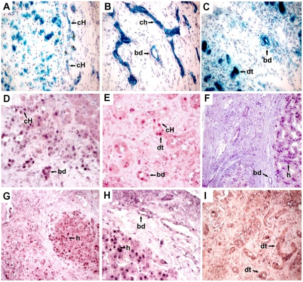
Methodology/principal findings: Livers of five children aged three years or less undergoing liver transplantation due to tyrosinemia (n = 1), biliary atresia (n = 2), or fulminant hepatitis (n = 2) were analyzed for senescence associated beta-galactosidase (SA-betagal) activity and p16INK4a, p21cip1 and p53. All livers displayed positive cellular staining for SA-betagal in the canals of Hering and interlobular biliary ducts. In the presence of cirrhosis (3/5 cases) SA-betagal was found at the cholangioles and hepatocytes surrounding the regenerative nodules. Children with fulminant hepatic failure without cirrhosis had significant ductular transformation with intense SA-betagal activity. No SA-betagal activity was evident in the fibrous septa. Staining for p53 had a similar distribution to that observed for SA-betagal. Staining for p16(INK4a) and p21(cip1) was positive in the explanted liver of the patient with tyrosinemia, in the hepatocytes, the canals of Hering, cholangioles and interlobular bile ducts. In the livers with fulminant hepatitis, p21(cip1) staining occurred in the areas of ductular transformation and in the interlobular bile ducts.
Conclusions/significance: Cellular senescence in livers of children with end stage disease is associated with damage rather than corresponding to an age dependent phenomenon. Further studies are needed to support the hypothesis that these senescence markers correlate with disease progression.
Conflict of interest statement
Figures
Similar articles
-
Increased expression of senescence-associated cell cycle regulators in the progression of biliary atresia: an immunohistochemical study.Histopathology. 2018 Jun;72(7):1164-1171. doi: 10.1111/his.13476. Epub 2018 Mar 9. Histopathology. 2018. PMID: 29392752
-
Telomere shortening in the damaged small bile ducts in primary biliary cirrhosis reflects ongoing cellular senescence.Hepatology. 2008 Jul;48(1):186-95. doi: 10.1002/hep.22348. Hepatology. 2008. PMID: 18536059
-
Bile ductular cells undergoing cellular senescence increase in chronic liver diseases along with fibrous progression.Am J Clin Pathol. 2010 Feb;133(2):212-23. doi: 10.1309/AJCPWMX47TREYWZG. Am J Clin Pathol. 2010. PMID: 20093230
-
Autophagy may precede cellular senescence of bile ductular cells in ductular reaction in primary biliary cirrhosis.Dig Dis Sci. 2012 Mar;57(3):660-6. doi: 10.1007/s10620-011-1929-y. Epub 2011 Oct 12. Dig Dis Sci. 2012. PMID: 21989821
-
Tumor suppressors and oncogenes in cellular senescence.Exp Gerontol. 2000 May;35(3):317-29. doi: 10.1016/s0531-5565(00)00083-8. Exp Gerontol. 2000. PMID: 10832053 Review.
Cited by
-
Knockout of l-Histidine Decarboxylase Prevents Cholangiocyte Damage and Hepatic Fibrosis in Mice Subjected to High-Fat Diet Feeding via Disrupted Histamine/Leptin Signaling.Am J Pathol. 2018 Mar;188(3):600-615. doi: 10.1016/j.ajpath.2017.11.016. Epub 2017 Dec 15. Am J Pathol. 2018. PMID: 29248461 Free PMC article.
-
Immunomodulatory Role of the Extracellular Matrix Within the Liver Disease Microenvironment.Front Immunol. 2020 Nov 11;11:574276. doi: 10.3389/fimmu.2020.574276. eCollection 2020. Front Immunol. 2020. PMID: 33262757 Free PMC article. Review.
-
Hepatic senescence, the good and the bad.World J Gastroenterol. 2019 Sep 14;25(34):5069-5081. doi: 10.3748/wjg.v25.i34.5069. World J Gastroenterol. 2019. PMID: 31558857 Free PMC article. Review.
-
Transient c-Src Suppression During Endodermal Commitment of Human Induced Pluripotent Stem Cells Results in Abnormal Profibrotic Cholangiocyte-Like Cells.Stem Cells. 2019 Mar;37(3):306-317. doi: 10.1002/stem.2950. Epub 2018 Dec 17. Stem Cells. 2019. PMID: 30471152 Free PMC article.
-
Cellular Senescence in Hepatocellular Carcinoma: The Passenger or the Driver?Cells. 2022 Dec 29;12(1):132. doi: 10.3390/cells12010132. Cells. 2022. PMID: 36611926 Free PMC article. Review.
References
-
- Hayflick L, Moorhead PS. The serial cultivation of human diploid cell strains. Exp Cell Res. 1961;25:585–621. - PubMed
-
- Maier A, Westendorp R, Van Heemst D. Beta-galactosidase activity as a biomarker of replicative senescence during the course of human fibroblast cultures. Ann N Y Acad Sci. 2007;1100:323–32. - PubMed
-
- Zhang H. Molecular signaling and genetic pathways of senescence: Its role in tumorigenesis and aging. J Cell Physiol. 2007;210:567–574. - PubMed
-
- Campisi J. Cancer, aging and cellular senescence. In Vivo. 2000;14:183–188. - PubMed
Publication types
MeSH terms
Substances
LinkOut - more resources
Full Text Sources
Other Literature Sources
Research Materials
Miscellaneous
