

MMP-9 gene ablation and TIMP-4 mitigate PAR-1-mediated cardiomyocyte dysfunction: a plausible role of dicer and miRNA
- PMID: 20422465
- PMCID: PMC2921878
- DOI: 10.1007/s12013-010-9084-1
MMP-9 gene ablation and TIMP-4 mitigate PAR-1-mediated cardiomyocyte dysfunction: a plausible role of dicer and miRNA
Abstract
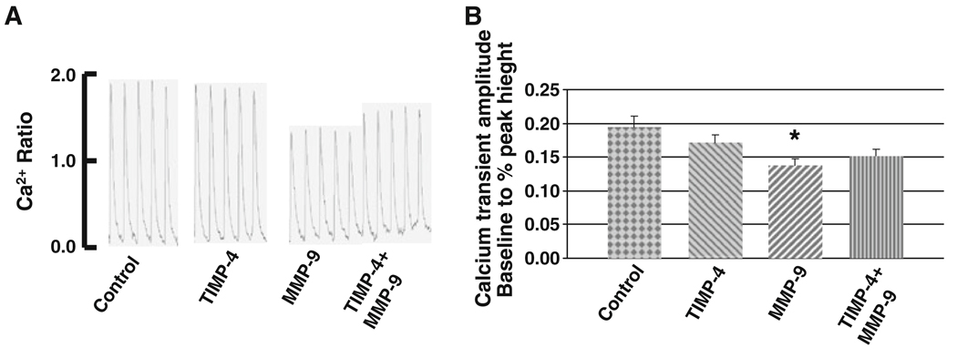
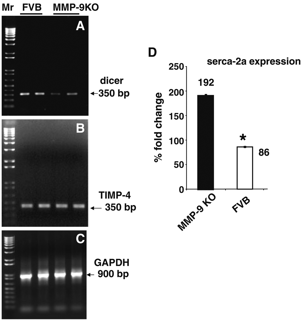
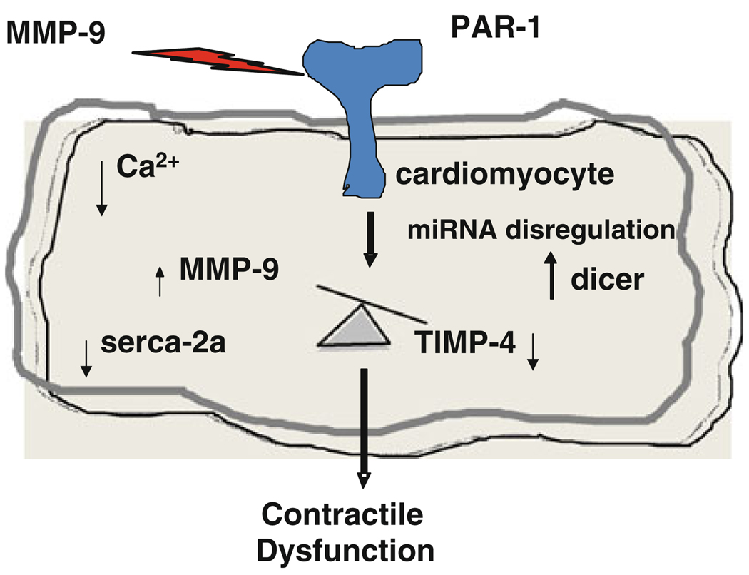
Although matrix metalloproteinase-9 (MMP-9) is involved in cardiomyocytes contractility dysfunction, tissue inhibitor of metalloproteinase-4 (TIMP-4) mitigates the effect of MMP-9, and proteinase-activated receptor-1 (PAR-1, a G-protein couple receptor, GPCR) is involved in the signaling cascade of MMP-9-mediated cardiac dysfunction, the mechanism(s) are unclear. To test the hypothesis that induction of dicer and differential expression of microRNAs (miRNAs) contribute, in part, to the down regulation of sarcoplasmic reticulum calcium ATPase isoform 2a (serca-2a) in MMP-9 and PAR-1-mediated myocytes dysfunction, ventricular cardiomyocytes were isolated from C57BL/6J mice and treated with 3 ng/ml of MMP-9, 12 ng/ml of TIMP-4, and 10 and 100 microM of PAR-1 antagonist with MMP-9. Specific role of MMP-9 was determined by using MMP-9 knock out (MMP-9KO) and their corresponding control (FVB) mice. Ion Optics video-edge detection system and Fura 2-AM loading were used for determining the contractility and calcium release from cardiomyocytes. Quantitative and semi-quantitative PCR were used to determine the expression of dicer, TIMP-4 and serca-2a. miRNA microarrays were used for assessing the expression of different miRNAs between MMP-9KO and FVB cardiomyocytes. The results suggest that MMP-9 treatment attenuates the voltage-induced contraction of primary cardiomyocytes while TIMP-4, an inhibitor of MMP-9, reverses the inhibition. MMP-9 treatment is also associated with reduced Ca(2+) transients. This effect is blocked by a PAR-1 antagonist, suggesting that PAR-1 mediates this effect. The effect is not as great at high concentrations (100 microM) perhaps due to mild toxicity. The PAR-1 antagonist effect did not affect calcium transients unlike TIMP-4. Interestingly, we show that MMP-KO myocytes contract more rapidly and release more Ca(2+) than FVB. The relevant RNA species serca-2a is induced and dicer is inhibited. There is selective inhibition of miR-376b and over-expression of miR-1, miR-26a, miR-30d, and miR-181c in MMP-9KO that are implicated in regulation of G-PCR and calcium handling.
Figures


Similar articles
-
Cardiac matrix: a clue for future therapy.Biochim Biophys Acta. 2013 Dec;1832(12):2271-6. doi: 10.1016/j.bbadis.2013.09.004. Epub 2013 Sep 17. Biochim Biophys Acta. 2013. PMID: 24055000 Free PMC article. Review.
-
MicroRNAs are involved in homocysteine-induced cardiac remodeling.Cell Biochem Biophys. 2009;55(3):153-62. doi: 10.1007/s12013-009-9063-6. Epub 2009 Aug 11. Cell Biochem Biophys. 2009. PMID: 19669742 Free PMC article.
-
Differential expression of dicer, miRNAs, and inflammatory markers in diabetic Ins2+/- Akita hearts.Cell Biochem Biophys. 2014 Jan;68(1):25-35. doi: 10.1007/s12013-013-9679-4. Cell Biochem Biophys. 2014. PMID: 23797610 Free PMC article.
-
Role of matrix metalloproteinase-9 in endothelial apoptosis in chronic heart failure in mice.J Appl Physiol (1985). 2005 Dec;99(6):2398-405. doi: 10.1152/japplphysiol.00442.2005. Epub 2005 Aug 4. J Appl Physiol (1985). 2005. PMID: 16081621
-
MicroRNAs are essential for stretch-induced vascular smooth muscle contractile differentiation via microRNA (miR)-145-dependent expression of L-type calcium channels.J Biol Chem. 2012 Jun 1;287(23):19199-206. doi: 10.1074/jbc.M112.341073. Epub 2012 Apr 2. J Biol Chem. 2012. PMID: 22474293 Free PMC article.
Cited by
-
The Biology and Function of Tissue Inhibitor of Metalloproteinase 2 in the Lungs.Pulm Med. 2022 Dec 31;2022:3632764. doi: 10.1155/2022/3632764. eCollection 2022. Pulm Med. 2022. PMID: 36624735 Free PMC article. Review.
-
Cardiac matrix: a clue for future therapy.Biochim Biophys Acta. 2013 Dec;1832(12):2271-6. doi: 10.1016/j.bbadis.2013.09.004. Epub 2013 Sep 17. Biochim Biophys Acta. 2013. PMID: 24055000 Free PMC article. Review.
-
Sensory Neuropathy Affects Cardiac miRNA Expression Network Targeting IGF-1, SLC2a-12, EIF-4e, and ULK-2 mRNAs.Int J Mol Sci. 2019 Feb 25;20(4):991. doi: 10.3390/ijms20040991. Int J Mol Sci. 2019. PMID: 30823517 Free PMC article.
-
Fibrosis-Related Gene Expression in Single Ventricle Heart Disease.J Pediatr. 2017 Dec;191:82-90.e2. doi: 10.1016/j.jpeds.2017.08.055. Epub 2017 Oct 16. J Pediatr. 2017. PMID: 29050751 Free PMC article.
-
A follow-up study for left ventricular mass on chromosome 12p11 identifies potential candidate genes.BMC Med Genet. 2011 Jul 26;12:100. doi: 10.1186/1471-2350-12-100. BMC Med Genet. 2011. PMID: 21791083 Free PMC article.
References
-
- Tyagi SC, Hoit BD. Metalloproteinase in myocardial adaptation and maladaptation. Journal of Cardiovascular Pharmacology and Therapeutics. 2002;7:241–246. - PubMed
-
- Ali MA, Schulz R. Activation of MMP-2 as a key event in oxidative stress injury to the heart. Frontiers in Bioscience. 2009;14:699–716. - PubMed
-
- Macfarlane SR, Seatter MJ, Kanke T, Hunter GD, Plevin R. Proteinase-activated receptors. Pharmacological Reviews. 2001;53:245–282. - PubMed
-
- Sabri A, Muske G, Zhang H, Pak E, Darrow A, Andrade-Gordon P, et al. Signaling properties and functions of two distinct cardiomyocyte protease-activated receptors. Circulation Research. 2000;86:1054–1061. - PubMed
Publication types
MeSH terms
Substances
Grants and funding
LinkOut - more resources
Full Text Sources
Research Materials
Miscellaneous
