Review
. 2010 Apr;55(4):308-16.
doi: 10.1097/fjc.0b013e3181d89670.

Vascular oxidative stress: the common link in hypertensive and diabetic vascular disease

Affiliations
Review

Vascular oxidative stress: the common link in hypertensive and diabetic vascular disease

Richard A Cohen et al. J Cardiovasc Pharmacol. 2010 Apr.

Abstract

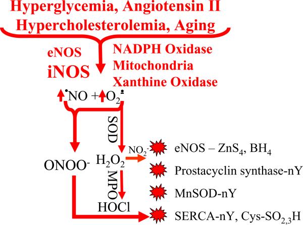
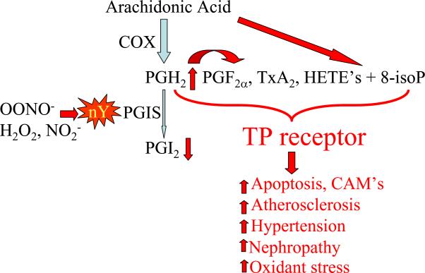
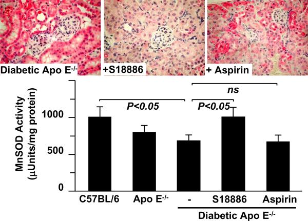
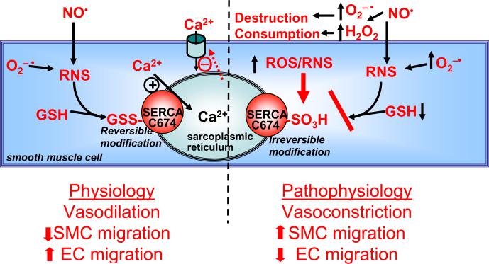
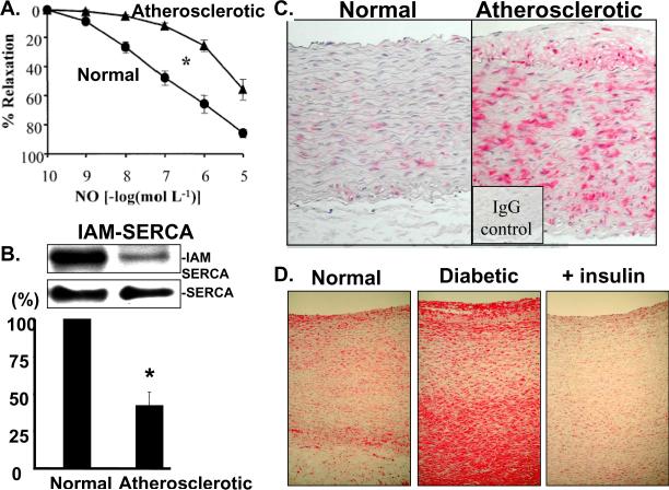
Vascular disease in hypertension and diabetes is associated with increased oxidants. The oxidants arise from NADPH oxidase, xanthine oxidase, and mitochondria. Superoxide anion and hydrogen peroxide are produced by both leukocytes and vascular cells. Nitric oxide is produced in excess by inducible nitric oxide synthase, and the potent oxidant, peroxynitrite, is formed from superoxide and nitric oxide. The damage to proteins caused by oxidants is selective, affecting specific oxidant-sensitive amino acid residues. With some important vascular proteins, for example, endothelial nitric oxide synthase, prostacycline synthase, and superoxide dismutase, oxidation of a single susceptible amino acid inactivates the enzyme. The beneficial effects of antioxidants, at least in animal models of hypertension and diabetes, can in part be ascribed to protection of these and other proteins. Mutant proteins lacking their reactive constituent can recapitulate some disease phenotypes suggesting a pathogenic role of the oxidation. Thus, many of the shared functional abnormalities of hypertensive and diabetic blood vessels may be caused by oxidants. Although studies using antioxidants have failed in patients, the successful treatment of vascular disease with HMG-CoA reductase inhibitors, thromboxane A2 antagonists, and polyphenols may depend on their anti-inflammatory effects and ability to decrease production of damaging oxidants.


Figures

Figure 1
Figure 1
Cardiovascular risk factors increase oxidants and protein oxidation. Major cardiovascular risk factors increase vascular production of nitric oxide (•NO) and superoxide anion (O2−•) by increasing the expression and/or activity of endothelial and inducible •NO synthase (eNOS, iNOS), NADPH oxidase, xanthine oxidase, as well as increasing production of mitochondrial O2−•. •NO and O2−• react rapidly to form peroxynitrite anion (OONO) which can increase tyrosine nitration (nY), cysteine (Cys) and zinc thiolate (ZnS4) oxidation (SO2,3H). Superoxide dismutases (SOD) form H2O2 which can also oxidize proteins, or together with leukocyte myeloperoxidase (MPO) can form hypochlorous acid (HOCl) and with nitrite (NO2) can form nY on proteins. Important cardiovascular proteins are affected including eNOS, prostacyclin synthase, MnSOD, the sarco-endoplasmic reticulum calcium ATPase (SERCA).
Figure 2
Figure 2
Tyrosine nitration (nY) of prostacyclin synthase (PGIS) increases stimulation of thromboxane A2 (TP) receptors. Nitration of PGIS at tyrosine-430 inactivates the enzyme resulting in shunting of arachidonic acid metabolites to products that stimulate the TP receptor. Cyclooxygenase produces prostaglandin endoperoxide (PGH2) which produces more prostaglandin (F) and thromboxane (Tx) A2. More arachidonic acid derived hydroxyeicosatetraenoic acids (HETE's) also are produced. Furthermore, oxidants generate more 8-isoprostanes (isoP) directly from arachidonic acid which also stimulates TP receptors. These products can all be implicated in apoptotic and inflammatory cell responses, increased atherosclerosis, hypertension, and nephropathy. TP receptor stimulation also further augments the generation of reactive oxygen species.
Figure 3
Figure 3
Tyrosine nitration and inactivation of MnSOD in diabetic apoliprotein E deficient mice is prevented by TP antagonist. Upper panels show examples of immunohistochemical staining of kidneys of atherosclerotic, hyperlipidemic apolipoprotein E deficient mice that were given type-1 diabetes by administration of streptozotocin. The red staining was achieved with a sequence-specific antibody that detects nitration of tyrosine-34 of MnSOD. Treatment of the mice with the TP antagonist, S18886 restored staining to a level indistinguishable from that in non-diabetic control mice, whereas aspirin had no significant effect. The bar graph shows that the renal MnSOD enzymatic activity was significantly decreased from control in the diabetic mice, and that treatment with S18886, but not aspirin prevented the decrease. Data from reference 64.
Figure 4
Figure 4
Redox regulation of SERCA by reactive oxygen/nitrogen species. RNS produced by •NO and O2−• increase glutathione adducts (GSS-) of cysteine(C)-674 of SERCA. This increases Ca2+ uptake into sarcoplasmic reticulum stores, inhibiting store-dependent Ca2+ influx, and decreasing cytosolic Ca2+ which causes vasodilation, inhibits smooth muscle cell (SMC) migration, and increases endothelial cell (EC) migration. Under pathophysiological conditions higher levels of ROS increase the destruction and consumption of •NO, producing RNS which can oxidize the SERCA C674 thiol (−SO3H), preventing its reversible S-glutathiolation and blocking the stimulation of SERCA by •NO. Thus, the redox status of C674 can determine physiological and pathophysiological changes in vascular tone and cell migration. From reference 8.
Figure 5
Figure 5
Oxidation of SERCA in atherosclerotic and diabetic aorta. A. Impaired aortic relaxations to •NO in rabbits made atherosclerotic by feeding a high cholesterol diet for 10 weeks. B. The decreased response can be explained in part by decreased labeling of the free thiol on cysteine-674 with biotin-tagged IAM (upper blot) summarized in bar graph. There is no change in total SERCA expression (lower blot). C and D. Immunohistochemical staining of oxidized SERCA cysteine-674 by a sequence specific antibody that recognizes the sulfonic acid thiol. Increased staining is seen in atherosclerotic rabbit aorta (C) obtained from the same study as data in panels A and B, as well as in the aorta of a pig maintained diabetic and hypercholesterolemic for 1 year, but not in a diabetic pig treated with insulin for 1 year. Data from references 8 and.

References

    1. Contreras F, Rivera M, Vasquez J, De la Parte MA, Velasco M. Diabetes and hypertension physiopathology and therapeutics. J Hum Hypertens. 2000;14(Suppl 1):S26–S31. - PubMed
    1. Adler AI, Stratton IM, Neil HA, Yudkin JS, Matthews DR, Cull CA, Wright AD, Turner RC, Holman RR. Association of systolic blood pressure with macrovascular and microvascular complications of type 2 diabetes (UKPDS 36): prospective observational study. BMJ. 2000;321(7258):412–9. - PMC - PubMed
    1. Stamler J, Vaccaro O, Neaton JD, Wentworth D. Diabetes, other risk factors, and 12-yr cardiovascular mortality for men screened in the Multiple Risk Factor Intervention Trial. Diabetes Care. 1993;16(2):434–44. - PubMed
    1. Savoia C, Schiffrin EL. Inhibition of the renin angiotensin system: implications for the endothelium. Curr Diab Rep. 2006;6(4):274–8. - PubMed
    1. Adachi T, Schoneich C, Cohen RA. S-glutathiolation in redox-sensitive signaling. Drug Discov Today. 2005;2(1):39–46.

Publication types