. 2010 Apr;2(4):1206-10.
doi: 10.1021/am100045v.

Antibacterial nanoparticle monolayers prepared on chemically inert surfaces by cooperative electrostatic adsorption (CELA)

Affiliations

Antibacterial nanoparticle monolayers prepared on chemically inert surfaces by cooperative electrostatic adsorption (CELA)

Sabil Huda et al. ACS Appl Mater Interfaces. 2010 Apr.

Abstract

Cooperative electrostatic adsorption (CELA) is used to deposit monolayer coatings of silver nanoparticles on relatively chemically inert polymers, polypropylene, and Tygon. Medically relevant components (tubing, vials, syringes) coated by this method exhibit antibacterial properties over weeks to months with the coatings being stable under constant-flow conditions. Antibacterial properties of the coatings are due to a slow release of Ag(+) from the particles. The rate of this release is quantified by the dithiol-precipitation method coupled with inductively coupled plasma optical emission spectrometer (ICP-OES) analysis.


Figures

FIGURE 1
FIGURE 1
Cooperative electrostatic adsorption. (a) Negatively charged NPs do not adsorb onto oxidized surfaces presenting residual negative charge. (b) Similarly, positively charged NPs give only very sparse coatings (~5% surface coverage) due to the repulsions between adsorbed particles. (c) Mixtures of positively and negatively charged nanoparticles adsorb cooperatively and yield dense coatings (~70% surface coverage). Scale bar for (a)–(c) is 200 nm; in all cases, the NPs are deposited on silicon. (d) Coatings deposited on glass and composed of only silver NPs (e.g., AgMUA and AgTMA) appear orange-red; those comprising gold and silver particles (e.g., AuMUA and AgTMA) are violet. Scale bar in (d) represents 10 mm. Also see fig. S1 in the Supporting Information.
FIGURE 2
FIGURE 2
Examples of medically relevant components coated with bacteriostatic NP monolayers of oppositely charged nanoparticles. (a) Tygon-R tubing (scale bar = 16 mm) (b) Polypropylene micro-pipette tips (scale bar = 8 mm) (c) Glass vials (scale bar = 8 mm) (d) Polypropylene syringes (scale bar = 32 mm). Yellow-orange coatings are made of AgMUA/AgTMA; pink coatings, from AuMUA/AgTMA. Uncolored pieces are shown for reference. All coatings were deposited by immersion (~10 min) in aqueous NP solutions.
FIGURE 3
FIGURE 3
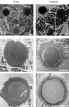
(a,b) Comparison of antibacterial properties of all-silver, AgMUA/AgTMA, and gold-silver, AuMUA/AuTMA, coatings deposited on polypropylene disks (all disk are 10 mm in diameter and are delineated by dashed lines). For both types of bacteria, E.Coli (left column) and S.Aureus (right column), the bacteria-free (clear) zones of inhibition are more pronounced for the all-silver coatings than for silver-gold coatings. Note the absence of inhibition around uncoated glass disks. (c,d) Zones of inhibition around pieces of PVC-based Tygon-R coated with AgMUA/AgTMA and tested on E.Coli (c) and on S.Aureus (d). (e, f) Zones of inhibition for Nylon coated with AgMUA/AgTMA tested on (e) E.coli and (f) S.Aureus. All scale bars (a–f) correspond to 5 mm. All images were taken 16 hrs after plating the bacteria.
FIGURE 4
FIGURE 4
(a) Scheme illustrating crosslinking of the deposited NP coatings with alkane dithiols (red arcs). (b) The middle images in each triad show as-prepared coatings (all-Ag on the left; mixed Ag/Au on the right) deposited on the inner surface of polypropylene syringes. Under flow of buffer (here, PBS, 1.3 mL/min), the coatings disintegrate overnight and the syringes loose the characteristic hue. When, however, the coatings are reinforced by crosslinking, the coatings remain stable under buffer flow for days to at least two weeks. Stability was confirmed by the lack of changes in the coating’s UV-Vis spectra. The scale bar corresponds to 17 mm.
FIGURE 5
FIGURE 5
(a) Scheme of the selective precipitation method in which addition of dithiols (red arcs = HS-(CH2)6-SH) causes crosslinking and aggregation of AgNPs (larger, gray circles) but not of Ag+ ions (smaller, brown circles). The concentration of ions released from the NPs and remaining in solution can be then determined by ICPOES. (b) Percentage of Ag+ cations released from AgNPs as a function of time. Error bars are based on the measurements of six samples for each time point. Measurements for each nanoparticles “leak” the cations at an approximately constant rate. Note that after four months, only ~3% of NP contents leaks out indicating that the NP coatings can provide long-term antibacterial protection.

References

    1. Son WK, Youk JH, Lee TS, Park WH. Macromol. Rapid Commun. 2004;25:1632–1637.
    1. Feng QL, Wu J, Chen GQ, Cui FZ, Kim TN, Kim JO. J. Biomed. Mater. Res. 2000;52:662–668. - PubMed
    1. Clement JL, Jarrett PS. Met. Based Drugs. 1994;1:467–482. - PMC - PubMed
    1. Pai MP, Pendland SL. Ann. Pharmacother. 2003;37:192–196. - PubMed
    1. Li Y, Leung P, Yao L, Song QW, Newton E. J. Hosp. Infect. 2006;62:58–63. - PubMed

Publication types