Review
. 2010;17(19):2059-69.
doi: 10.2174/092986710791233706.

Regulation of the protein C anticoagulant and antiinflammatory pathways

Affiliations
Review

Regulation of the protein C anticoagulant and antiinflammatory pathways

A R Rezaie. Curr Med Chem. 2010.

Abstract

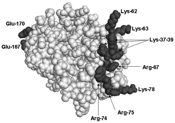
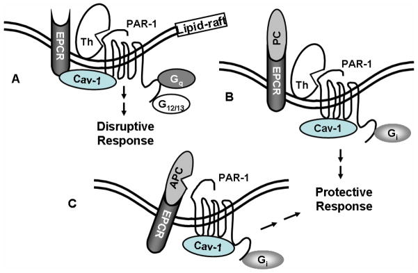
Protein C is a vitamin K-dependent anticoagulant serine protease zymogen in plasma which upon activation by the thrombin-thrombomodulin complex down-regulates the coagulation cascade by degrading cofactors Va and VIIIa by limited proteolysis. In addition to its anticoagulant function, activated protein C (APC) also binds to endothelial protein C receptor (EPCR) in lipid-rafts/caveolar compartments to activate protease- activated receptor 1 (PAR-1) thereby eliciting antiinflammatory and cytoprotective signaling responses in endothelial cells. These properties have led to FDA approval of recombinant APC as a therapeutic drug for severe sepsis. The mechanism by which APC selects its substrates in the anticoagulant and antiinflammatory pathways is not well understood. Recent structural and mutagenesis data have indicated that basic residues of three exposed surface loops known as 39-loop (Lys-37, Lys-38, and Lys-39), 60-loop (Lys-62, Lys- 63, and Arg-67), and 70-80-loop (Arg-74, Arg-75, and Lys-78) (chymotrypsin numbering) constitute an anion binding exosite in APC that interacts with the procoagulant cofactors Va and VIIIa in the anticoagulant pathway. Furthermore, two negatively charged residues on the opposite side of the active-site of APC on a helical structure have been demonstrated to determine the specificity of the PAR-1 recognition in the cytoprotective pathway. This article will review the mechanism by which APC exerts its proteolytic function in two physiologically inter-related pathways and how the structure- function insights into determinants of the specificity of APC interaction with its substrates in two pathways can be utilized to tinker with the structure of the molecule to obtain APC derivatives with potentially improved therapeutic profiles.


Figures

Fig. 1
Fig. 1
Crystal structure of the catalytic domain of APC. The side chains of basic residues of three loops (39, 60, and 70–80) and acidic residues of 162-helix (Glu-167 and Glu-170) are shown. The coordinates (Protein Data Bank code 1AUT) were used to prepare the figure [4].
Fig. 2
Fig. 2
Crystal structure of the catalytic domain of APC. (A) The three dimensional positions of Arg-67 and Asp-82 on two anti-parallel β structures of APC are shown. (B) Intra-atomic distances between NH1 guanidyl of Arg-67 and carboxylic oxygens of Asp-82.
Fig. 3
Fig. 3
Hypothetical model of protein C activation by the thrombin-TM complex. (A) Arg-67 and Asp-82 are involved in electrostatic interactions in the zymogen protein C in the absence of Ca2+. The activation peptide (AP) of protein C is not complimentary to fit optimally into the active-site pocket of thrombin in the absence of Ca2+. (B) The binding of Ca2+ to the 70-80-loop of protein C disrupts the salt-bridge/hydrogen bond between Arg-67 and Asp-82, thereby relocating Arg-67 to an inhibitory position for interaction with thrombin in the absence of TM. The metal ion also induces conformational change in the activation peptide of protein C, which is still not complimentary for the active-site pocket of free thrombin. (C) The acidic EGF5 domain of TM binds to exosite-1 of thrombin and the acidic EGF4 domain binds to the basic exosite of the Ca2+-stabilized protein C (including Arg-67), thereby altering the conformation of the active-site and/or the extended binding pocket residues of thrombin (including Arg-35), thus facilitating the docking of the activation peptide of protein C in the catalytic groove of thrombin (see the text for further details).
Fig. 4
Fig. 4
Cartoons of wild-type and mutant prothrombin activation by factor Xa. (A) The proteolytic cleavage of prothrombin at Arg-320 by factor Xa yields an activation intermediate that is an active product called meizothrombin. A second cleavage at Arg-271 by factor Xa is required to separate the catalytic domain of prothrombin from the non-catalytic domain (Gla, Kringle-1 and Kringle-2 domains) to yield thrombin. Further cleavages can occur at Arg-155 and Arg-286 in a feed-back reaction by both thrombin and meizothrombin. (B) The substitution of the Gla domain of prothrombin with the corresponding domain of protein C and its Arg-155, Arg-271 and Arg-286 residues with 3 Ala’s yields a mutant of prothrombin (3A-prothrombin/PC-Gla) which can be activated by factor Xa through the cleavage of Arg-320 to yield meizothrombin/PC-Gla which cannot be further processed by either factor Xa or the resulting mutant meizothrombin.
Fig. 5
Fig. 5
Cartoons of PAR-1 activation by APC and thrombin when EPCR is either free or occupied by its ligand protein C. (A) EPCR is associated with caveolin-1 (Cav-1) in the lipid-rafts of endothelial cells when the receptor is not occupied by the Gla domain of protein C/APC. Thrombin cleavage of PAR-1 elicits disruptive signaling responses through coupling the receptor to G12/13 and Gq proteins. (B) The occupancy of EPCR by protein C (PC) results in the dissociation of EPCR from caveolin-1, thereby switching the specificity of PAR-1 signaling by coupling it to the Gi protein. Thus, thrombin cleavage of PAR-1 initiates protective response when EPCR is occupied. (C) The same as (B) except that the EPCR and PAR-1 dependent protective signaling response is elicited by APC.

References

    1. Esmon CT. Molecular events that control the protein C anticoagulant pathway. Thromb Haemost. 1993;70:1–5. - PubMed
    1. Stenflo J. Structure-function relationships of epidermal growth factor modules in vitamin K-dependent clotting factors. Blood. 1991;78:1637–1651. - PubMed
    1. Walker FJ, Fay PJ. Regulation of blood coagulation by the protein C system. FASEB J. 1992;6:2561–2567. - PubMed
    1. Mather T, Oganessyan V, Hof P, Huber R, Foundling S, Esmon C, Bode W. The 2.8 Å crystal structure of Gla-domainless activated protein C. EMBO J. 1996;15:6822–6831. - PMC - PubMed
    1. Dahlbäck B. Protein S and C4b-binding protein: Components involved in the regulation of the protein C anticoagulant system. Thromb Haemostas. 1991;66:49–61. - PubMed

Publication types

MeSH terms