. 2010 May;160(2):220-32.
doi: 10.1111/j.1476-5381.2010.00698.x.

Isoflurane protects cardiomyocytes and mitochondria by immediate and cytosol-independent action at reperfusion

Affiliations

Isoflurane protects cardiomyocytes and mitochondria by immediate and cytosol-independent action at reperfusion

D Pravdic et al. Br J Pharmacol. 2010 May.

Abstract

Background and purpose: The volatile anaesthetic isoflurane protects the heart from ischaemia and reperfusion (I/R) injury when applied at the onset of reperfusion [anaesthetic postconditioning (APoC)]. However, the mechanism of APoC-mediated protection is unknown. In this study, we examined the effect of APoC on mitochondrial bioenergetics, mitochondrial matrix pH (pH(m)) and cytosolic pH (pH(i)), and intracellular Ca(2+).

Experimental approach: Cardiac mitochondria from Wistar rats were isolated after in vivo I/R with or without APoC (1.4%-vol isoflurane, 1 minimum alveolar concentration), and mitochondrial permeability transition pore (mPTP) opening, mitochondrial membrane potential (DeltaPsi(m)), and oxygen consumption were assessed. In isolated cardiomyocytes and isolated mitochondria I/R injury was produced in vitro, with or without APoC (0.5 mM isoflurane). Intracellular Ca(2+), pH(m), pH(i) and DeltaPsi(m) were monitored with SNARF-1, TMRE and fluo-4, respectively. Myocyte survival was assessed when APoC was induced at pH 7.4 and 7.8. In isolated mitochondria oxygen consumption and ATP synthesis were measured.

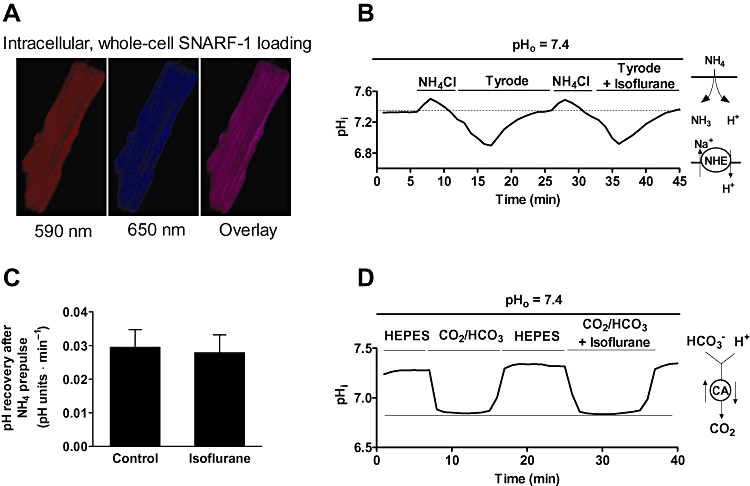
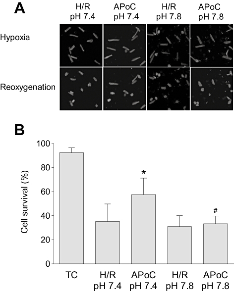
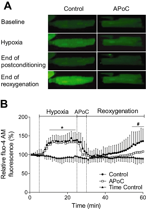
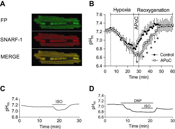
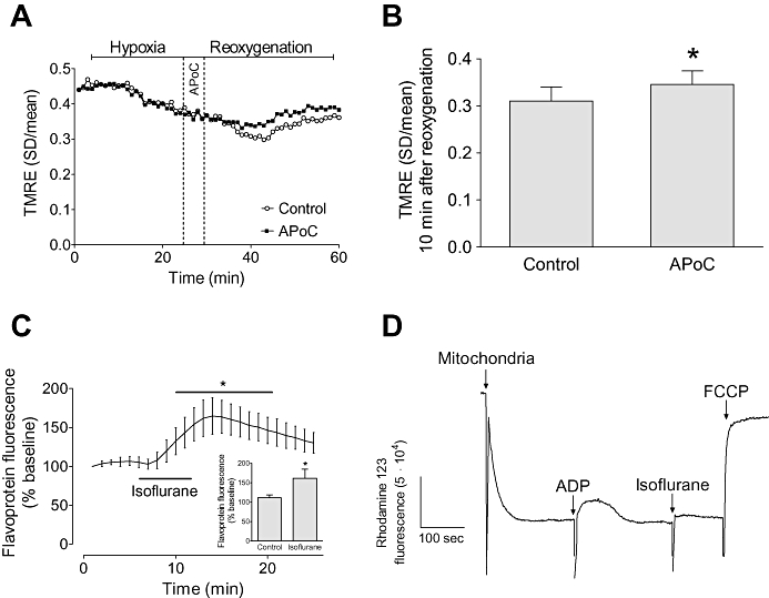
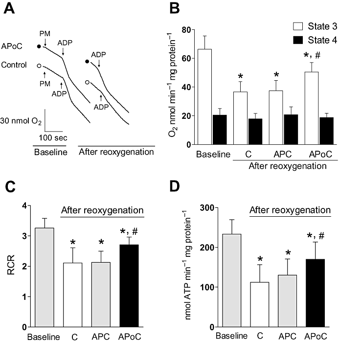
Key results: In vivo APoC protected against mPTP opening, slowed mitochondrial respiration and depolarized mitochondria. APoC decreased the number of hypercontracted cardiomyocytes at pH 7.4, but not at pH 7.8. APoC attenuated intracellular Ca(2+) accumulation, maintained lower pH(m), and preserved DeltaPsi(m) during reoxygenation. Isoflurane did not affect the regulation of cytosolic pH. In mitochondria, APoC preserved ATP production rate and respiration.

Conclusions and implications: At reperfusion, APoC inhibited mitochondrial respiration, depolarized mitochondria and acidified pH(m). These events may lead to inhibition of mPTP opening and, consequently, to preserved DeltaPsi(m) and ATP synthesis. This reduces intracellular Ca(2+) overload and cell death.


Figures

Figure 5
Figure 5
Isoflurane does not modulate NHE exchanger and carbonic anhydrase activities. (A) Representative images showing whole-cell loading of SNARF-1. (B) Representative trace of pHi recovery from the cardiomyocyte following exposure to NH4Cl (10 mM) in 4-(2-hydroxyethyl)-1-piperazineethanesulfonic acid (HEPES)-buffered Tyrode solution, pHo 7.4. The trace demonstrates that after two consecutive NH4Cl pulses, pHi recovery from an intracellular acid load was not different in the presence of isoflurane. (C) Summary for recovery of pHi from NH4Cl -induced acidosis in the presence and subsequent absence of 0.5 mM isoflurane. (D) Effect of isoflurane on carbonic anhydrase activity following intracellular acid loading induced by application of 5% CO2/HCO-3. The magnitude of acidosis was not different in the presence of isoflurane. Data are presented as means ± standard deviation. Traces are representative from at least five measurements. SNARF-1, 5-(and-6)-carboxy SNARF-1, acetoxymethyl ester; NHE, Na+-H+ exchanger, CA, carbonic anhydrase.
Figure 1
Figure 1
Isoflurane postconditioning in vivo protects mitochondria from ischaemia and reperfusion (I/R) injury. (A) Original recordings of ΔΨm in mitochondria from control and APoC hearts in the presence of pyruvate/malate. (B) Summarized data showing ΔΨm relative to maximum depolarization. (C) The amount of Ca2+ necessary to open the mPTP in sham, control and APoC mitochondria. Values are means ± standard deviation. *P < 0.05 versus sham, #P < 0.05 versus control, n= 9. ΔΨm, mitochondrial membrane potential; mPTP, mitochondrial permeability transition pore; FCCP, carbonyl cyanide 4-(trifluoromethoxy)phenylhydrazone; S2, state 2 respiration; APoC, isoflurane postconditioning.
Figure 2
Figure 2
Isoflurane postconditioning protects isolated cardiomyocytes from H/R injury. (A) Representative images of isolated cardiomyocyte changes during H/R. During reoxygenation after hypoxia cardiomyocytes became spherical, hypercontracted and exhibited membrane blebs. APoC protected cardiomyocytes against H/R injury at pH 7.4, but the protection was lost when APoC was performed at pH 7.8. (B) Summary graph showing the percentage of cell survival after 35 min of reoxygenation. Data are means ± standard deviation; *P < 0.05 versus time control; #P < 0.05 versus ApoC; n= 5. TC, time control; H/R, hypoxia and reoxygenation; APoC, isoflurane postconditioning.
Figure 3
Figure 3
APoC decreases cytosolic Ca2+ overload in cardiomyocytes during reoxygenation. (A) Representative image of isolated cardiomyocytes loaded with fluo-4, fluorescent indicator for intracellular Ca2+. Under hypoxic conditions intracellular Ca2+ increased relative to pre-hypoxic values and then recovered at reoxygenation. During reoxygenation, Ca2+ remained low at early reoxygenation, but then increased. APoC decreased cytosolic Ca2+ overload during reoxygenation. (B) Summarized data for intracellular Ca2+ during hypoxia and reoxygenation. Standard deviations (SDs) are omitted for clarity in APoC group. Values are means ± SD; *P < 0.05 versus time control; #P < 0.05 versus control; n= 17. APoC, isoflurane postconditioning.
Figure 4
Figure 4
APoC prevents rapid pHm recovery in cardiomyocytes at reoxygenation. (A) Representative images showing selective mitochondrial loading of SNARF-1. SNARF-1 colocalizes with mitochondrial flavoproteins (FP). (B) pHm decreased during hypoxia as shown by a decrease in SNARF-1 ratio. Upon reoxygenation, pHm quickly recoverd to baseline levels. Treatment of cardiomyocytes with isoflurane at the beginning of reoxygenation preserved acidic intracellular pHm during reoxygenation as compared with the control group. (C) The panel shows representative time lapse of baseline pHm recording. Baseline pHm was reduced upon addition of isoflurane into superfusing solution, and this effect was reversible after anaesthetic washout. (D) Administration of the mitochondrial uncoupler 2-4-dinitrophenol (DNP) prevented any further effect of isoflurane on pHm. Data are presented as means ± standard deviation, *P < 0.05 versus APoC; n= 20. APoC, isoflurane postconditioning; SNARF-1, 5-(and-6)-carboxy SNARF-1, acetoxymethyl ester.
Figure 6
Figure 6
APoC preserves ΔΨm in isolated myocytes throughout reoxygenation. (A) Representative recording of ΔΨm changes during H/R. Hypoxia caused a significant decrease in the standard deviation of TMRE fluorescence indicating mitochondrial depolarization whereas reoxygenation caused an increase in the standard deviation indicating recovery of ΔΨm. Treatment of cardiomyocytes with isoflurane at the beginning of reoxygenation preserved ΔΨm as compared with control cells. (B) Summary data for control and APoC groups at 10 min of reoxygenation. (C) Effect of isoflurane on mitochondrial flavoprotein fluorescence, and summary data (inset). (D) Representative trace of ΔΨm in isolated mitochondria. Isoflurane induced slight depolarization of ΔΨm. Data are means ± standard deviation; n= 15; n= 6 in flavoprotein and isolated mitochondria experiments. *P < 0.05 versus control. APoC, isoflurane postconditioning, TMRE, tetramethylrhodamine ethyl ester. FCCP, carbonyl cyanide 4-(trifluoromethoxy)phenylhydrazone.
Figure 7
Figure 7
In vitro APoC protects isolated mitochondria from H/R injury. (A) Typical recordings showing respiration under baseline condition and after reoxygenation in control and isoflurane-postconditioned mitochondria. Oxygen consumption was initiated by addition of pyruvate/malate (PM), accelerated by addition of ADP (state 3 respiration), and decelerated after all ADP was consumed (state 4 respiration). Following H/R, state 3 respiration was preserved in APoC-treated mitochondria. (B) After H/R, state 3 respiration decreased in all groups; however, state 3 respiration in APoC-treated mitochondria was better preserved as compared with control and preconditioned mitochondria. (C) Respiratory control ratio (RCR) was better maintained in APoC treated mitochondria, whereas preconditioning did not improve RCR. (D) The rate of ATP production was decelerated after reoxygenation. However, ATP synthesis in APoC-treated mitochondria was better preserved as compared with control and preconditioned mitochondria. Values are mean ± SD. *P < 0.05 versus baseline or APoC; #P < 0.05 versus control or APC. C, control; APoC, isoflurane postconditioning; APC, isoflurane preconditioning; open circle, control; solid circle, isoflurane postconditioning, n= 7 per group.

Comment in

References

    1. Abad MFC, Di Benedetto G, Magalhaes PJ, Filippin L, Pozzan T. Mitochondrial pH monitored by a new engineered green fluorescent protein mutant. J Biol Chem. 2004;279:11521–11529. - PubMed
    1. Aizawa K, Turner LA, Weihrauch D, Bosnjak ZJ, Kwok WM. Protein kinase C-epsilon primes the cardiac sarcolemmal adenosine triphosphate-sensitive potassium channel to modulation by isoflurane. Anesthesiology. 2004;101:381–389. - PubMed
    1. Alexander SPH, Mathie A, Peters JA. Guide to Receptors and Channels (GRAC), 4th edn. Br J Pharmacol. 2009;158(Suppl 1):S1–S254. - PMC - PubMed
    1. Brennan JP, Berry RG, Baghai M, Duchen MR, Shattock MJ. FCCP is cardioprotective at concentrations that cause mitochondrial oxidation without detectable depolarisation. Cardiovasc Res. 2006;72:322–330. - PubMed
    1. Buckler KJ, Vaughan-Jones RD. Application of a new pH-sensitive fluoroprobe (carboxy-SNARF-1) for intracellular pH measurement in small, isolated cells. Pflugers Arch. 1990;417:234–239. - PubMed

Publication types

MeSH terms