. 2010 May;160(2):233-45.
doi: 10.1111/j.1476-5381.2010.00663.x.

Methylenedioxymethamphetamine inhibits mitochondrial complex I activity in mice: a possible mechanism underlying neurotoxicity

Affiliations

Methylenedioxymethamphetamine inhibits mitochondrial complex I activity in mice: a possible mechanism underlying neurotoxicity

Elena Puerta et al. Br J Pharmacol. 2010 May.

Abstract

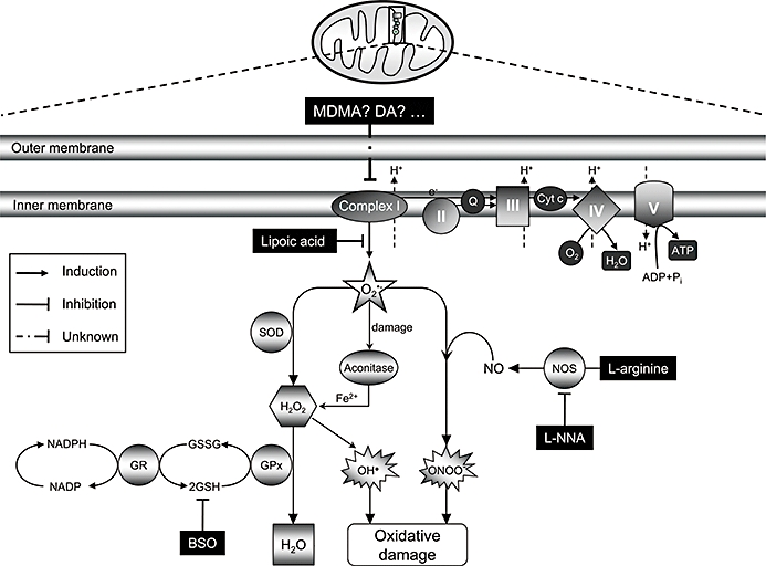
Background and purpose: 3,4-methylenedioxymethamphetamine (MDMA) causes a persistent loss of dopaminergic cell bodies in the substantia nigra of mice. Current evidence indicates that such neurotoxicity is due to oxidative stress but the source of free radicals remains unknown. Inhibition of mitochondrial electron transport chain complexes by MDMA was assessed as a possible source.

Experimental approach: Activities of mitochondrial complexes after MDMA were evaluated spectrophotometrically. In situ visualization of superoxide production in the striatum was assessed by ethidium fluorescence and striatal dopamine levels were determined by HPLC as an index of dopaminergic toxicity.

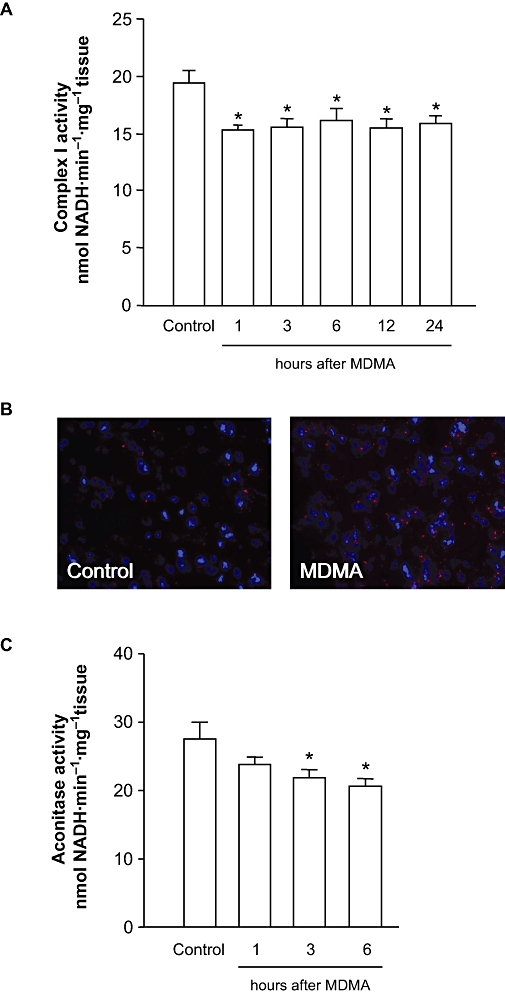
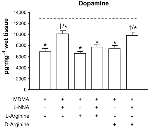
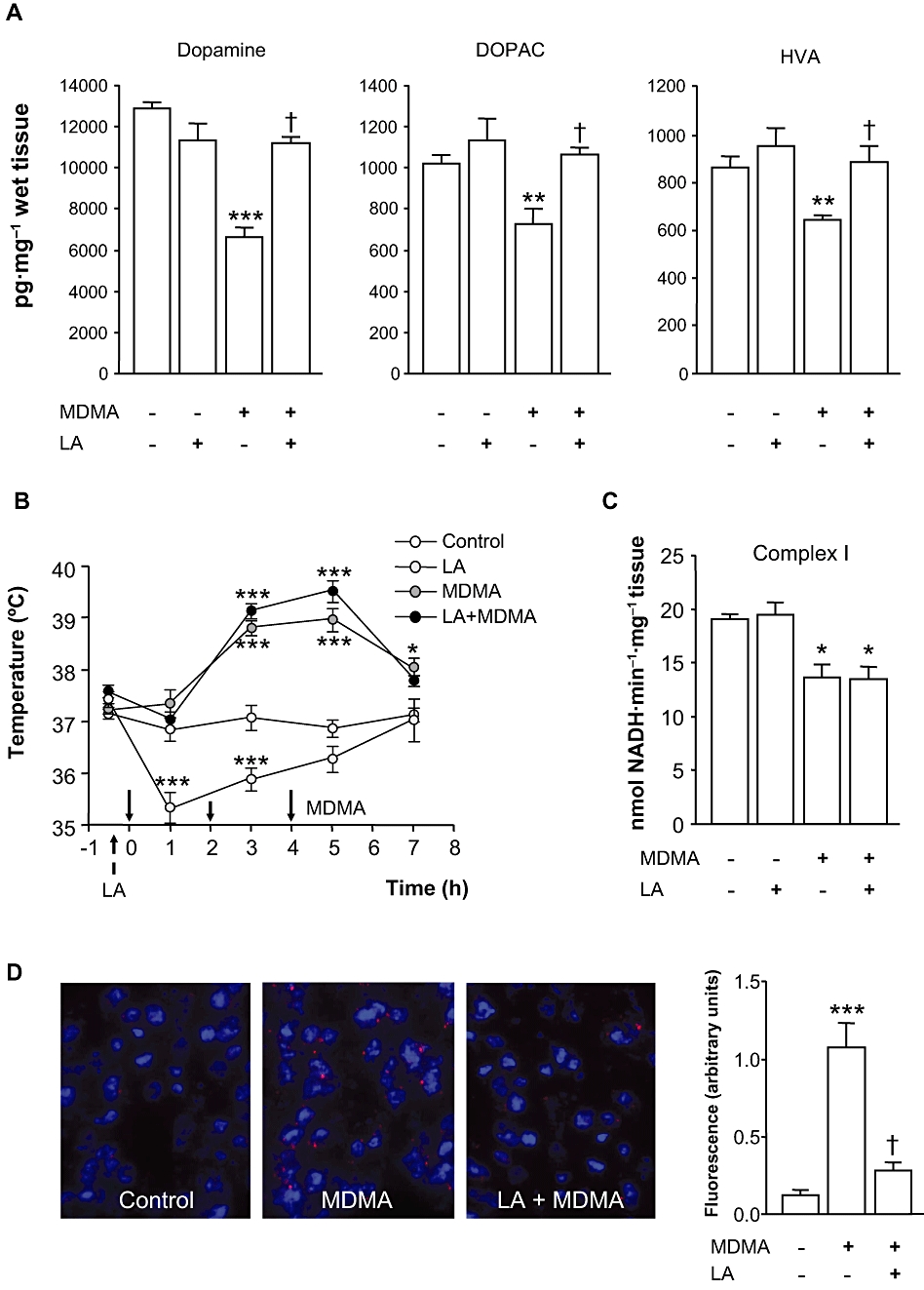
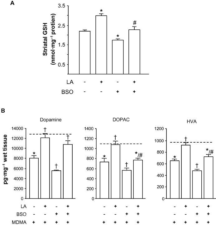
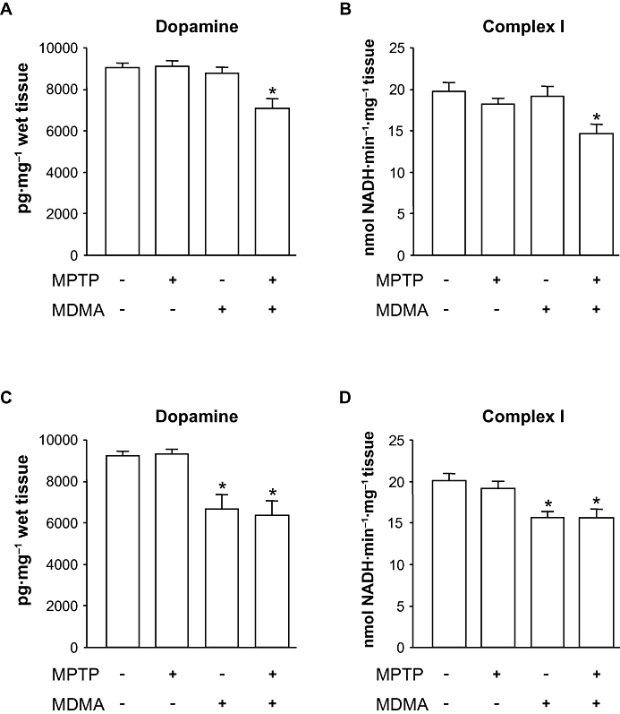
Key results: 3,4-methylenedioxymethamphetamine decreased mitochondrial complex I activity in the striatum of mice, an effect accompanied by an increased production of superoxide radicals and the inhibition of endogenous aconitase. alpha-Lipoic acid prevented superoxide generation and long-term toxicity independent of any effect on complex I inhibition. These effects of alpha-lipoic acid were also associated with a significant increase of striatal glutathione levels. The relevance of glutathione was supported by reducing striatal glutathione content with L-buthionine-(S,R)-sulfoximine, which exacerbated MDMA-induced dopamine deficits, effects suppressed by alpha-lipoic acid. The nitric oxide synthase inhibitor, N(G)-nitro-L-arginine, partially prevented MDMA-induced dopamine depletions, an effect reversed by L-arginine but not D-arginine. Finally, a direct relationship between mitochondrial complex I inhibition and long-term dopamine depletions was found in animals treated with MDMA in combination with 1-methyl-4-phenyl-1,2,3,6-tetrahydropyridine.

Conclusions and implications: Inhibition of mitochondrial complex I following MDMA could be the source of free radicals responsible for oxidative stress and the consequent neurotoxicity of this drug in mice.


Figures

Figure 1
Figure 1
Effect of MDMA on mitochondrial complex I and aconitase activities and superoxide (O2-) production. (A) Mitochondrial complex I in the striatum of mice killed 1, 3, 6, 12 and 24 h after MDMA (10, 20, 30 mg·kg−1 i.p. every 2 h). (B) Representative photomicrographs showing fluorescent ethidium signals 3 h after saline or MDMA. Note that MDMA treatment resulted in a significant increased in oxidized hydroethidine signals compared with saline-treated mice. (C) Effect of MDMA on aconitase activity in mice killed 1, 3 and 6 h after MDMA. The results shown as mean ± SEM (n= 10; A and C). Differences from saline: *P < 0.05. MDMA, 3,4-methylenedioxymethamphetamine.
Figure 2
Figure 2
Effect of the NOS inhibitor L-NNA alone or in combination with D- or L-arginine on MDMA-induced striatal dopamine loss. L-NNA (10 mg·kg−1 i.p.) or saline were administered 30 min before MDMA (10, 20, 30 mg·kg−1 i.p. every 2 h), and D- or L-arginine (300 mg·kg−1 i.p.) were given 5 min before L-NNA or saline. Mice were killed 7 days later. L-arginine but not D-arginine reversed the protection afforded by L-NNA against MDMA-induced dopamine depletions. Discontinous line (----) represents dopamine levels in saline-treated mice. Results shown as means ± SEM (n= 8–12). *P < 0.05 different from saline; †P < 0.05 different from MDMA-treated mice. L-NNA, NG-nitro-L-arginine; MDMA, 3,4-methylenedioxymethamphetamine; NOS, nitric oxide synthase.
Figure 3
Figure 3
Effect of LA on MDMA-induced dopaminergic deficits, mitochondrial complex I inhibition and ROS production. LA (100 mg·kg−1 i.p.) or vehicle were given twice daily for 2 days, and MDMA (10, 20, 30 mg·kg−1 i.p. 2 h apart) was given 30 min after the fourth administration of LA or vehicle. (A) Striatal dopamine, DOPAC and HVA levels of mice 7 days after MDMA given alone or in combination with LA. Results shown as means ± SEM in pg·mg−1 wet tissue (n= 8–10). (B) Effect of LA on MDMA-induced acute hyperthermia. Arrows denote administration of LA or MDMA (solid arrows). Mice temperatures were recorded at baseline (t=–30 min) and 1 h after every injection of MDMA up to 7 h. Values are means ± SEM (n= 8–12 mice per group). (C) Complex I activity measured in the striata of mice 3 h after treatments. Results shown as means ± SEM (n= 8–12). (D) Effect of LA on MDMA-induced O2•− production. O2•− radicals were measured in animals treated with saline (control) or MDMA alone or in combination with LA. Note that LA decreased the ethidium signal evoked by MDMA. Results shown as means ± SEM (n= 3–4). In all panels: *P < 0.05, **P < 0.01 or ***P < 0.001 versus control group; †P < 0.01, different from MDMA-treated animals. One-way anova followed by Newman–Keuls test. DOPAC, 3,4-dihydroxyphenylacetic acid; HVA, homovanillic acid; LA, α-lipoic acid; MDMA, 3,4-methylenedioxymethamphetamine; ROS, reactive oxygen species.
Figure 4
Figure 4
Effect of BSO on MDMA induced-toxicity. BSO (3 mmol·kg−1 i.p.) was given 24 h before the fourth injection of LA (4 × 100 mg·kg−1 i.p. 12 h apart). (A) Striatal GSH levels 24 h after BSO given alone or in combination with LA. Results shown as means ± SEM (n= 8–10). *P < 0.05 versus saline; #P < 0.05 versus BSO-treated animals. (B) Striatal dopamine, DOPAC and HVA levels of mice 7 days after administration of BSO or LA in combination with MDMA. Discontinous line (----) shows dopamine, DOPAC and HVA levels of saline-treated mice. Results shown as means ± SEM (n= 8–12). *P < 0.05, different from saline; †P < 0.05, different from MDMA-treated mice; #P < 0.05, different from BSO + MDMA treated animals. One-way anova followed by Newman–Keuls test. BSO, L-buthionine-(S,R)-sulfoximine; DOPAC, 3,4-dihydroxyphenylacetic acid; GSH, glutathione; LA, α-lipoic acid; MDMA, 3,4-methylenedioxymethamphetamine.
Figure 5
Figure 5
Effect of MPTP on MDMA-induced toxicity. (A) Striatal dopamine levels 7 days after the administration of MDMA (3 × 10 mg·kg−1 i.p. every 2 h) alone or in combination with MPTP (3 × 10 mg·kg−1 i.p. every 2 h). (B) mitochondrial complex I activity 3 h after the same drug treatments shown in panel A. (C) Striatal dopamine levels 7 days after the administration of MDMA (10, 20, 30 mg·kg−1 i.p. every 2 h) alone or in combination with MPTP (3 × 10 mg·kg−1 i.p. every 2 h). (D) mitochondrial complex I activity 3 h after the same drug treatments shown in panel C. Results shown as means ± SEM (n= 8–12). *P < 0.05 versus saline-treated mice. One-way anova followed by Newman–Keuls test. MDMA, 3,4-methylenedioxymethamphetamine; MPTP, 1-methyl-4-phenyl-1,2,3,6-tetrahydropyridine.
Figure 6
Figure 6
A hypothetical model of MDMA-induced dopaminergic toxicity in mice. The inhibition of mitochondrial complex I activity after systemic administration of MDMA promotes the generation of superoxide radicals (O2•−), which could lead to the formation of peroxynitrite (ONOO-), in the presence of NO, or to H2O2 by means of superoxide dismutase (SOD). The [4Fe-4S]2+ cluster of aconitase could then be inactivated by O2•−, peroxynitrite or H2O2 leading to the generation of Fe2+, which, together with H2O2, may increase the formation of hydroxyl radicals (OH) by the Fenton reaction. Glutathione (GSH) is involved both as a non-enzymic, free radical scavenger for ROS and/or peroxynitrite and enzymically to inactivate H2O2. In summary, inhibition of mitochondrial complex I activity could be a plausible source of free radicals responsible for oxidative damage to dopamine neurons caused by MDMA in mice. Further studies are needed, however, to resolve which is/are the specific compound(s) responsible for such effects. GPx, gluthathione peroxidase; GR, glutathione reductase; MDMA, 3,4-methylenedioxymethamphetamine; ROS, reactive oxygen species.

Comment in

References

    1. Aguirre N, Barrionuevo M, Ramírez MJ, Del Río J, Lasheras B. Alpha-lipoic acid prevents 3,4-methylenedioxy-methamphetamine (MDMA)-induced neurotoxicity. Neuroreport. 1999;10:3675–3680. - PubMed
    1. Albers DS, Zeevalk GD, Sonsalla PK. Damage to dopaminergic nerve terminals in mice by combined treatment of intrastriatal malonate with systemic methamphetamine or MPTP. Brain Res. 1996;718:217–220. - PubMed
    1. Aleardi AM, Benard G, Augereau O, Malgat M, Talbot JC, Mazat JP, et al. Gradual alteration of mitochondrial structure and function by beta-amyloids: importance of membrane viscosity changes, energy deprivation, reactive oxygen species production, and cytochrome c release. J Bioenerg Biomembr. 2005;37:207–225. - PubMed
    1. Alexander SPH, Mathie A, Peters JA. Guide to Receptors and Channels (GRAC), 4th edn. Br J Pharmacol. 2009;158(Suppl 1):S1–S254. - PMC - PubMed
    1. Alves E, Summavielle T, Alves CJ, Custódio JB, Fernandes E, de Lourdes Bastos M, et al. Ecstasy-induced oxidative stress to adolescent rat brain mitochondria in vivo: influence of monoamine oxidase type A. Addict Biol. 2009;14:185–193. - PubMed

Publication types

MeSH terms