. 2010 May;160(2):376-88.
doi: 10.1111/j.1476-5381.2010.00696.x.

Shikonin reduces oedema induced by phorbol ester by interfering with IkappaBalpha degradation thus inhibiting translocation of NF-kappaB to the nucleus

Affiliations

Shikonin reduces oedema induced by phorbol ester by interfering with IkappaBalpha degradation thus inhibiting translocation of NF-kappaB to the nucleus

I Andújar et al. Br J Pharmacol. 2010 May.

Abstract

Background and purpose: In the present paper we studied the effect of shikonin on ear oedema induced by 12-O-tetradecanoylphorbol-13-acetate (TPA), and determined the mechanisms through which shikonin might exert its topical anti-inflammatory action.

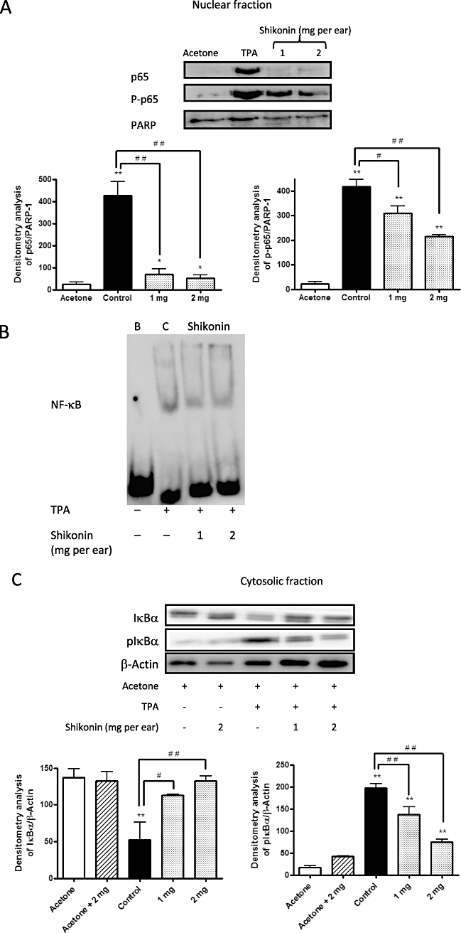
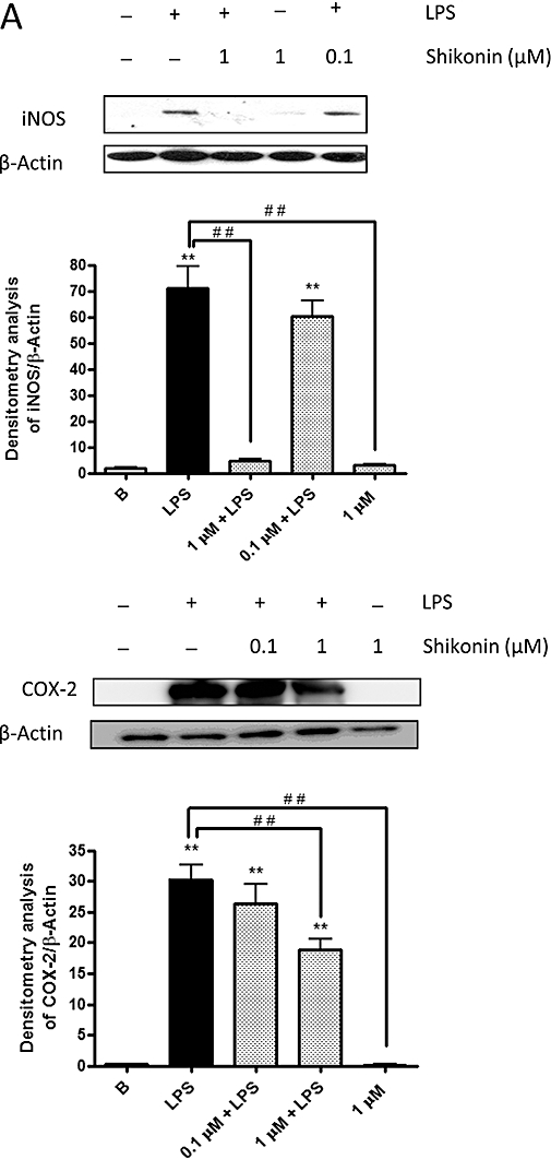
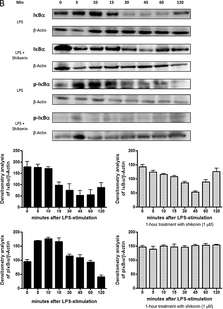
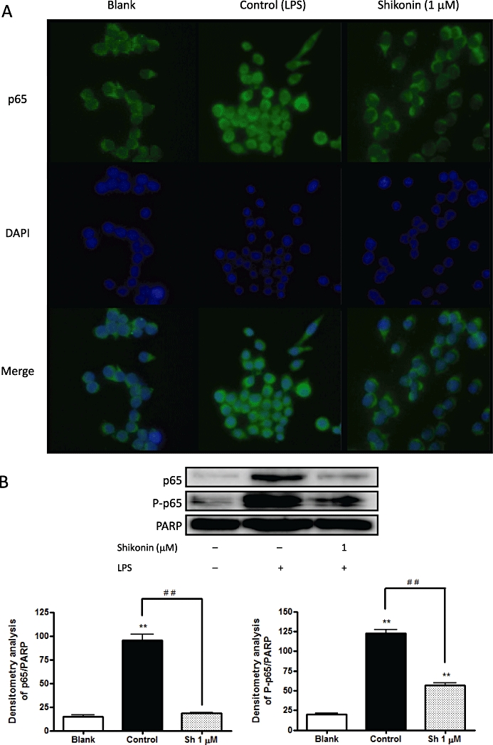
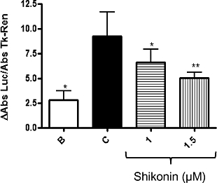
Experimental approach: Acute ear oedema was induced in mice by topical application of TPA. The in vitro assays used macrophages RAW 264.7 cells stimulated with lipopolysaccharide. Cyclooxygenase-2, inducible nitric oxide synthase, protein kinase Calpha, extracellular signal-regulated protein kinase (ERK), phosphorylated ERK (pERK), c-Jun N-terminal kinase (JNK), pJNK, p38, p-p38, p65, p-p65, inhibitor protein of nuclear factor-kappaB (NF-kappaB) (IkappaBalpha) and pIkappaBalpha were measured by Western blotting, activation and binding of NF-kappaB to DNA was detected by reporter gene and electrophoretic mobility shift assay, respectively, and NF-kappaB p65 localization was detected by immunocytochemistry.

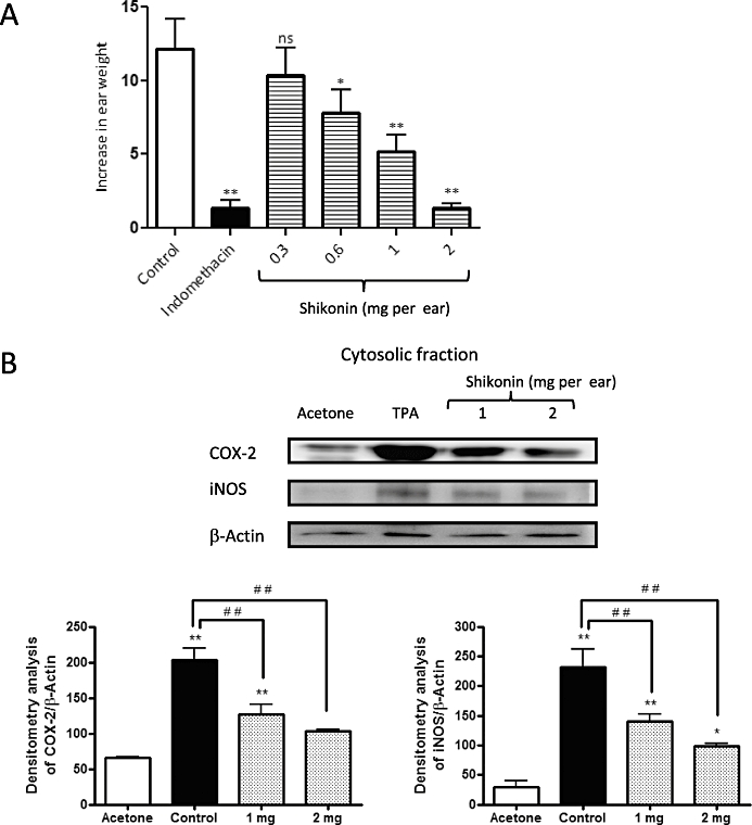
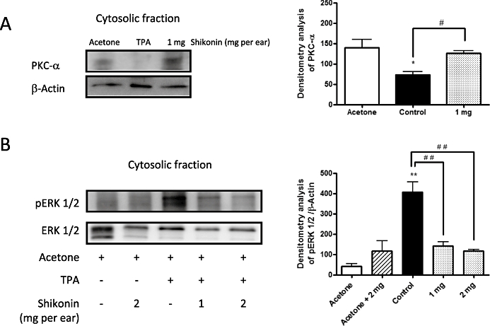
Key results: Shikonin reduced the oedema (inhibitory dose 50 = 1.0 mg per ear), the expression of cyclooxygenase-2 (70%) and of inducible nitric oxide synthase (100%) in vivo. It significantly decreased TPA-induced translocation of protein kinase Calpha, the phosphorylation and activation of ERK, the nuclear translocation of NF-kappaB and the TPA-induced NF-kappaB-DNA-binding activity in mouse skin. Moreover, in RAW 264.7 cells, shikonin significantly inhibited the binding of NF-kappaB to DNA in a dose-dependent manner and the nuclear translocation of p65.

Conclusions and implications: Shikonin exerted its topical anti-inflammatory action by interfering with the degradation of IkappaBalpha, thus inhibiting the activation of NF-kappaB.


Figures

Figure 1
Figure 1
Chemical structure of shikonin.
Figure 2
Figure 2
Inhibitory effects of shikonin (Sh) on 12-O-tetradecanoylphorbol-13-acetate (TPA)-induced mouse ear oedema. Mice topically treated with shikonin (0.3, 0.6, 1 and 2 mg per ear) were killed 1, 2 or 4 h after TPA application. (A) Oedema was measured after 4 h of TPA application; values are expressed as the increase in ear weight (ΔW) ± standard error of the mean, n= 6; *P < 0.05, **P < 0.01. ns, not significantly different; Dunnett's test. (B) Effect of shikonin treatment on levels of cyclooxigenase-2 and inducible nitric oxide synthase in ear tissue. The upper panels show an example of Western blot following probing with the corresponding antibody. The lower histograms represent the data derived from the Western blots following densitometry analysis. *P < 0.05, **P < 0.01, significantly different from the acetone group; #P < 0.05, ##P < 0.01, significantly different between the control group and the groups treated with shikonin; Dunnett's test. n= 3.
Figure 3
Figure 3
(A) Effect of shikonin on protein kinase Cα (PKC-α) enzymes in extracts of ear tissue after 12-O-tetradecanoylphorbol-13-acetate (TPA). (B) Effect of shikonin treatment on levels of phosphorylated and non-phosphorylated extracellular signal-regulated protein kinase (ERK 1/2) after 1 h of TPA application. The upper panels in Figures 3A,B show an example of Western blot following probing with the corresponding antibody. The lower histograms represent the data derived from the Western blots following densitometry analysis. *P < 0.05, **P < 0.01, significantly different from the acetone group; #P < 0.05, ##P < 0.01, significantly different between the control group and the groups treated with shikonin; Dunnett's test. n= 3–5.
Figure 4
Figure 4
( A) Nuclear cell extract prepared from the ears after 4 h of 12-O-tetradecanoylphorbol-13-acetate (TPA) application was analysed for p65 and P-p65. (B) Effect of shikonin on nuclear factor-κB (NF-κB) binding activity. B, blank (non-treated); C, control (TPA treated); Shikonin (TPA and shikonin treated). (C) Two hours after TPA application, the effect of shikonin treatment on levels of IκBα and pIκBα was assayed with the aid of Western blot techniques. The upper panels in Figures 4A,C show an example of Western blot following probing with the corresponding antibody. The lower histograms represent the data derived from the Western blots following densitometry analysis. *P < 0.05, **P < 0.01, significantly different from the acetone group; #P < 0.05, ##P < 0.01, significantly different between the control group and the groups treated with shikonin; Dunnett's test. n= 3–4.
Figure 5
Figure 5
(A) Effect of shikonin treatment on levels of inducible nitric oxide synthase and cyclooxygenase-2 in RAW 264.7 macrophages. The data given are the mean values ± standard error of the mean taken from six independent experiments for each enzyme. *P < 0.05, **P < 0.01, significantly different from the blank group; #P < 0.05, ##P < 0.01, significant difference between the control group and the groups treated with shikonin; Dunnett's test. (B) Shikonin inhibits lipopolysaccharide (LPS)-dependent IκBα degradation. The data given are the means ± standard error of the mean of values taken from four independent experiments. A representative experiment is shown in the upper panel. The lower histograms represent the data derived from the Western blots following densitometry analysis. In A and B, levels were normalised against β-actin.
Figure 5
Figure 5
(A) Effect of shikonin treatment on levels of inducible nitric oxide synthase and cyclooxygenase-2 in RAW 264.7 macrophages. The data given are the mean values ± standard error of the mean taken from six independent experiments for each enzyme. *P < 0.05, **P < 0.01, significantly different from the blank group; #P < 0.05, ##P < 0.01, significant difference between the control group and the groups treated with shikonin; Dunnett's test. (B) Shikonin inhibits lipopolysaccharide (LPS)-dependent IκBα degradation. The data given are the means ± standard error of the mean of values taken from four independent experiments. A representative experiment is shown in the upper panel. The lower histograms represent the data derived from the Western blots following densitometry analysis. In A and B, levels were normalised against β-actin.
Figure 6
Figure 6
(A) Shikonin suppresses p65 translocation to the nucleus in RAW 264.7 macrophages, as shown by an immunocytochemical analysis of p65 localization. p65 was indicated by the presence of fluorescent green, DNA by fluorescent blue. In non-stimulated cells (blank) and shikonin-treated RAW 264.7 cells, p65 was localised in the cytoplasm, whereas after treatment with lipopolysaccharide, p65 suffered nuclear translocation (control). (B) Shikonin inhibits lipopolysaccharide-induced nuclear translocation of p65 in RAW 264.7 macrophages. The upper panels show a representative Western blot following probing with the corresponding antibody. The bars represent the data derived from the Western blots following densitometry analysis. Levels were normalised against poly (ADP-ribose) polymerase-antibody. *P < 0.05, **P < 0.01; significantly different from the blank group; #P < 0.05, ##P < 0.01; significantly different between the control group and the groups treated with shikonin; Dunnett's test. n= 5.
Figure 7
Figure 7
Shikonin suppresses lipopolysaccharide (LPS)-induced NF-κB activity, as shown in the luciferase assay in RAW 264.7 macrophages. Luciferase activity was normalised to TK-Renilla activity. The data were obtained from four independent experiments and expressed as the mean (ΔAbs) ± standard error of the mean differences in absorbance of each group with the control group were determined by means of a one-way analysis of variance followed by Dunnett's test, n= 4; *P < 0.05, **P < 0.01.

References

    1. Afaq F, Saleem M, Aziz MH, Mukhtar H. Inhibition of 12-O-tetradecanoylphorbol-13-acetate-induced tumor promotion markers in CD-1 mouse skin by oleandrin. Toxicol Appl Pharmacol. 2004;195(3):361–369. - PubMed
    1. Baeuerle PA. IkB-NF-κB structures: at the interface of inflammation control. Cell. 1998;95:729–731. - PubMed
    1. Baeuerle PA, Baltimore D. NF-κB: ten years after. Cell. 1996;87:13–20. - PubMed
    1. Baggiolini M. Chemokines and leukocyte traffic. Nature. 1998;392:565–568. - PubMed
    1. Bell S, Degitz K, Quirling M, Jilg N, Page S, Brand K. Involvement of NF-κB signalling in skin physiology and disease. Cell Signal. 2003;15:1–7. - PubMed

Publication types

MeSH terms