. 2010 Sep;38(16):5366-83.
doi: 10.1093/nar/gkq285. Epub 2010 Apr 27.

CREB up-regulates long non-coding RNA, HULC expression through interaction with microRNA-372 in liver cancer

Affiliations

CREB up-regulates long non-coding RNA, HULC expression through interaction with microRNA-372 in liver cancer

Jiayi Wang et al. Nucleic Acids Res. 2010 Sep.

Abstract

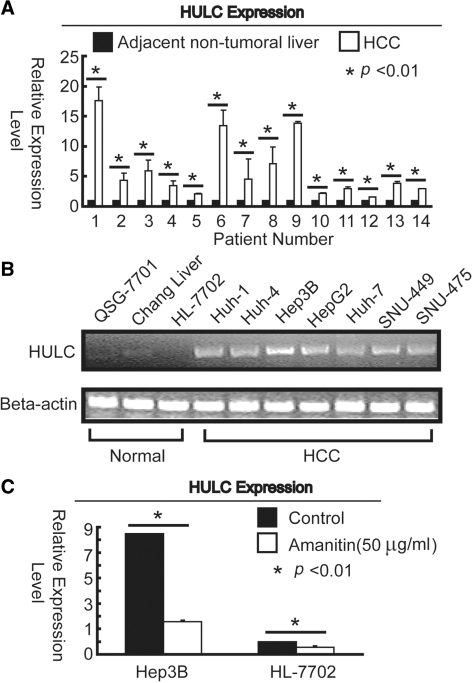
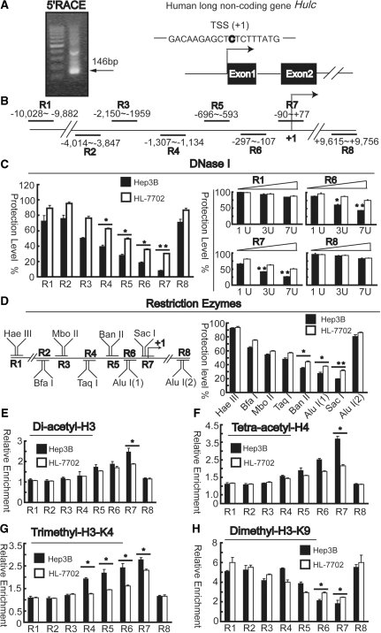
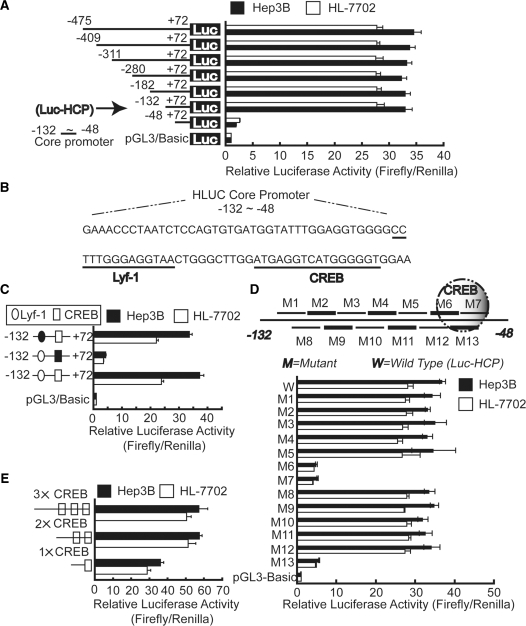
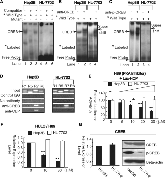
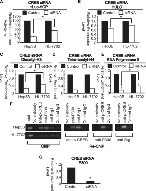
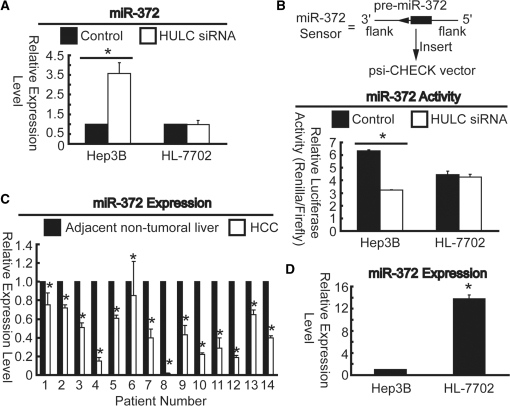
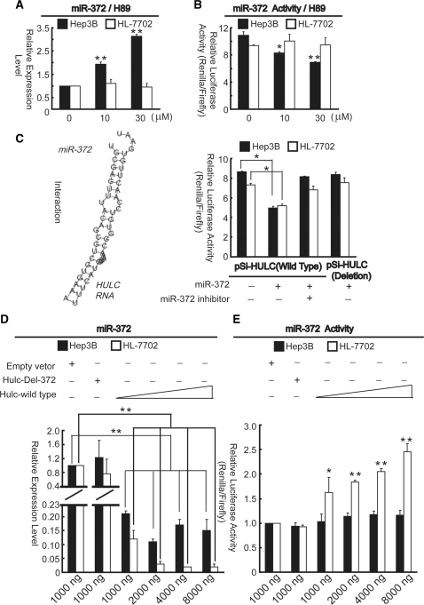
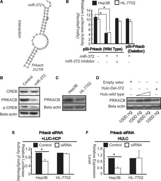
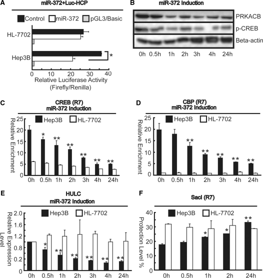
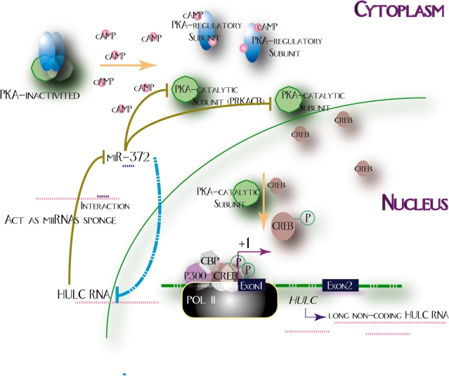
Long non-coding RNA (lncRNA), highly up-regulated in liver cancer (HULC) plays an important role in tumorigenesis. Depletion of HULC resulted in a significant deregulation of several genes involved in liver cancer. Although up-regulation of HULC expression in hepatocellular carcinoma has been reported, the molecular mechanisms remain unknown. In this study, we used in vivo and in vitro approaches to characterize cancer-dependent alterations in the chromatin organization and find a CREB binding site (encompassing from -67 to -53 nt) in the core promoter. Besides, we also provided evidence that PKA pathway may involved in up-regulation of HULC. Furthermore, we demonstrated HULC may act as an endogenous 'sponge', which down-regulates a series of microRNAs (miRNAs) activities, including miR-372. Inhibition of miR-372 leads to reducing translational repression of its target gene, PRKACB, which in turn induces phosphorylation of CREB. Over-expression of miR-372 decreases the association of CREB with the proximal promoter, followed by the dissociation of P300, resulting in a change of the histone 'code', such as in deacetylation and methylation. The study elucidates that fine tuning of HULC expression is part of an auto-regulatory loop in which it's inhibitory to expression and activity of miR-372 allows lncRNA up-regulated expression in liver cancer.


Figures

Figure 1.
Figure 1.
Up-regulation of HULC in HCC and liver cancer cell lines. (A) Change in HULC expression between adjacent normal and HCC tissues was quantified by real-time PCR. (B) HULC expression levels in different liver cell lines by semi-qRT-PCR. β-actin was treated as internal control. (C) Hep3B and HL-7702 cells were stimulated with α-amanitin (50 μg/ml) for 24 h. All results are means ± SD of three independent experiments. The asterisk indicates statistical significance at the P < 0.01 level using t-test.
Figure 2.
Figure 2.
Analysis of chromatin accessibility around the Hulc promoter. (A) 5′RACE PCR products suggest the putative TSS is ∼146 bp upstream of the HULC reverse 5′RACE primer (left panel). Mapping the TSS of Hulc in HCC (right panel). (B) Schematic representation of the DNA regions around Hulc promoter, which can be amplified by corresponding primer sets. (C) DNase I accessibility of Hulc gene in Hep3B and HL-7702 cells. Nuclei from two cell lines were harvested and treated with three units (left panel) or increasing amount (1–7 units, right panel) of DNase I for 5 min at room temperature. Then the genomic DNA was purified and quantitated relative to DNA from undigested nuclei using the primers described in (B) by quantitative PCR and listed as percent protected. (D) Restriction enzymes accessibility of Hulc gene in Hep3B and HL-7702 cells. Nuclei was treated with 25–150 units of restriction enzymes for 30 min at 37°C. Then the genomic DNA was purified and quantitated relative to DNA from undigested nuclei using the primers described in (B) by quantitative PCR and listed as percent protected. Chromatin from Hep3B and HL-7702 cells were harvested and precipitated with anti-diacetyl-H3 (E), anti-tetra-acetyl-H4 (F), anti-tri-methyl-H3-K4 (G) and anti-dimethyl-H3-K9 (H) antibodies. After DNA recovery, the precipitates were evaluated by real-time PCR for the level of enrichment over negative control antibody using primer sets described in (B). All results are the means ± SD of three independent experiments. The asterisk indicates statistical significance using t-test, *P < 0.05; **P < 0.01.
Figure 3.
Figure 3.
CREB-binding site contributes to the activation of the promoter. (A) Hep3B and HL-7702 cells were transiently co-transfected with reporter plasmids containing truncated versions of the promoter region of the Hulc gene, as indicated, and pRL-TK. Luc-HCP is defined as the reporter containing the shortest promoter region (from −132 to +72), which core elements located in. (B) Transcription factor binding sites present in the core promoter region (from −132 to −48) were predicated by web software TFSEARCH and MatInspector, as indicated by dashed lines. (C) Cells were transiently transfected with wild-type Luc-HCP as well as Luc-HCP which Lyf-1 or CREB binding site was deleted. Lyf-1-binding site is indicated by circle, whereas CREB-binding site is indicated by rectangle; deleted Lyf-1 and CREB sites are shown as black cycle and rectangle, respectively. (D) Scanning mutational analysis of the fragment from −132 to 48. As shown on the upper panel, a series of 8-bp substitutions were made within the fragment from −132 to 48 of the reporter construct Luc-HCP (from −132 to +72). M6, M7 and M13 overlap the possible CREB-binding site. (E) Wild-type Luc-HCP and Luc-HCP containing additional one (2×) or two (3×) possible CREB-binding sites were co-transfected with pRL-TK into Hep3B and HL-7702 cells. Firefly luciferase activity was normalized to Renilla luciferase activity, and the relative luciferase activities are presented as fold increase over the promoter-less pGL3 basic vector. Horizontal column lengths represent the means ± SD of three independent experiments.
Figure 4.
Figure 4.
HULC expression is regulated by phospho-CREB through PKA pathway in liver cancer cells. (A) Upon interaction with the nuclear extract of Hep3B (left panel) and HL-7702 (right panel), wild probe generated specific band (lanes 1 and 4) which was self-competed by 30-fold excess of unlabeled probe (lanes 2 and 5). No competition was observed when using the same amounts of mutant probe (lanes 3 and 6). (B) Interaction with anti-CREB antibody resulted in a super shift band formation in both two cell lines (lanes 2 and 4). (C) Interaction with anti-phospho-CREB antibody resulted in a super shift band formation (lane 2) in Hep3B, but not detected in HL-7702 cell line (lane 4). (D) ChIP analysis was performed to qualitative confirm the interaction of CREB and CBP with the Hulc promoter in vivo in Hep3B (upper panel) and HL-7702 (lower panel) cells using primer sets R1, R5, R7 and R8 described in Figure 2B. PCR products from the ChIP assay were run on an agarose gel. As the negative controls, the protein–DNA complexes were incubated without antibodies or with non-specific control IgG. The input DNA represents one-fifth of the starting material. (E) Pre-incubation with H89 was performed 1 day before transiently co-transfected Luc-HCP and internal control pRL-TK plasmids into Hep3B and HL-7702 cells and further incubation for 1 day in the continued presence of indicated amount of H89. Firefly luciferase activity was normalized to Renilla luciferase activity. Results are shown as relative percentage to those of cells untreated H89. (F) Total RNA was extracted for measurement of HULC mRNA expression level after treatment by indicated amount of H89 by real-time PCR. *P <0.05; **P <0.01 versus the corresponding untreated cells (E and F). (G) Endogenous CREB mRNA was quantified by real-time PCR and normalized to β-actin RNA in Hep3B and HL-7702 cells (left panel). The whole lysates of Hep3B and HL-7702 cells were examined by immunoblotting with antibodies against CREB, p-CREB and β-actin (right panel).
Figure 5.
Figure 5.
Reduction of CREB results in changes of chromatin accessibility and histone acetylation. (A) RNAi for CREB reduced Hulc promoter activity in Hep3B and HL-7702 cells. CREB or negative control siRNAs were co-transfected with Luc-HCP into cells. Luciferase assays were performed 24 h after transfection. Firefly luciferase activity was normalized to Renilla luciferase activity. (B) Endogenous HULC mRNA was quantified by real-time PCR and normalized to β-actin RNA. Chromatin from Hep3B and HL-7702 cells treated with control or CREB specific siRNA was harvested and precipitated with anti-diacetyl-H3 (C), anti-tetra-acetyl-H4 (D) and anti-pol II (E) antibodies. After DNA recovery, the precipitates were evaluated by real-time PCR for the level of enrichment over negative control antibody. (F) ChIP and re-ChIP experiments performed with anti-phosph-CREB, anti-P300 and anti-Brg I antibodies on Hep3B and HL-7702 cells. (G) Chromatin from Hep3B cells treated with control or CREB specific siRNA was harvested and precipitated with anti-P300 antibody. All the results (A–E and G) are the means of three independent experiments ± SD. Results of cells transfected siRNA against CREB are normalized to those of cells treated by negative control siRNA. The asterisk indicates statistical significance at the P < 0.05 level using t-test.
Figure 6.
Figure 6.
HULC inhibits miR-372 expression and activity. (A) Expression profiling of miR-372 48 h after HULC siRNA treatment in Hep3B and HL-7702 cells. (B) Construction of miR-372 sensor (upper panel). Genomic sequences (∼200 bp) flanking pre-miRNAs were reverse inserted into the psiCHECK vector, placing the 3′UTR with the miRNA binding sites downstream of coding sequence of Renilla luciferase. Luciferase assays indicated that enhanced miRNAs’ activities lead to decreasing levels of Renilla luciferase after treatment by HULC siRNA in Hep3B and HL-7702 cells (lower panel). Renilla luciferase activities were normalized to firefly luciferase activities. *P < 0.01, versus the cells treated with negative control siRNA (A and B). (C) Change in miR-372 expression between adjacent normal and HCC tissues was quantified by real-time PCR. *P < 0.01, versus adjacent normal tissue. (D) Differences in miR-372 expression level between Hep3B and HL-7702 cell lines. *P < 0.01, versus Hep3B cells. All the results described above are means of three independent experiments ± SD.
Figure 7.
Figure 7.
Interaction between HULC RNA and miR-372. (A) Relative fold change of miR-372 in the presence of indicated amount of H89 for 24 h. (B) Pre-incubation with H89 was performed 1 day before transiently transfected miR-372 sensor into Hep3B and HL-7702 cells and further incubation for 1 day in the continued presence of indicated amount of H89. *P < 0.05 and **P < 0.01, versus the cells untreated with H89 (A and B). (C) Sequence inspection by microInspector online software reveals HULC RNA contains a conserved element complementary to miR-372 (left panel). psiCHECK vectors containing HULC RNA with (pSi-HULC, wild-type) or without (pSi-HULC, deletion) predicted miR-372 binding site were co-transfected with miR-372 or both miR-372 and miR-372 inhibitor simultaneously (right panel). Luciferase assays were performed 24 h post-transfection. *P < 0.05. (D) The indicated amount of HULC-pcDNA3.1(+) with or without miR-372 binding site and empty vectors were transfected into Hep3B and HL-7702 cells grown on a six-well plate for 48 h. Then the total RNA was extracted for detection of miR-372. **P < 0.01. (E) The miR-372 sensor reporter plasmid was co-transfected with pcDNA3.1(+) vector as described in (D) and then cells were harvested, lysed, centrifuged and the pellet subjected to luciferase assay. *P < 0.05 and **P < 0.01, versus cells transfected with empty vectors. Renilla luciferase activity was normalized to Firefly luciferase activity (C and E). All results are shown as means of three independent experiments ± SD.
Figure 8.
Figure 8.
Interaction between Prkacb and miR-372. (A) Prkacb 3′UTR contains a miR-372 target site, as shown in the left panel, and confirmed by Luciferase assays (right panel). pSi-Prkacb contains Prkacb 3′UTR. Experimental procedure is similar with that described in Figure 7C. (B) Immunoblotting assay indicated that over-expression of miR-372 (50 nM) results in down-regulation of PRKACB, thus reduces phosphorylation of CREB in Hep3B cells. (C) Differences in PRKACB expression level between Hep3B and HL-7702 cell lines were measured by immunoblotting assay. (D) PRKACB expression level was detected by immunoblotting assay after transfection with pcDNA3.1(+) vector as indicated in HL-7702 cells. (E) Prkacb or negative control siRNAs were co-transfected with Luc-HCP into Hep3B and HL-7702 cells. Luciferase assays were performed 24 h after transfection. Firefly luciferase activities were normalized to Renilla luciferase activities. (F) Endogenous HULC mRNA was quantified by real-time PCR and normalized to β-actin RNA. Results of RNAi are shown as fold change to the control. *P < 0.01 versus corresponding control cells. All results are shown as means of three independent experiments ± SD.
Figure 9.
Figure 9.
Over-expression of miR-372 inhibits Hulc promoter activity. (A) Luciferase assays indicated that miR-372 could reduce promoter activity of Luc-HCP in Hep3B cells compared to that of HL-7702 cells. Firefly luciferase activity was normalized to Renilla luciferase activity, and the relative luciferase activities are presented as fold increase over the promoter-less pGL3-basic vector. *P < 0.01 versus control cells. (B) Immunoblotting assays indicated that over-expression of miR-372 could gradually down-regulation of PRKACB, thus reduce phosphorylation of CREB in Hep3B cells. Chromatin from Hep3B and HL-7702 cells was harvested 0, 0.5, 1, 2, 3, 4 and 24 h and precipitated with anti-CREB (C) and anti-CBP (D) antibodies. After DNA recovery, the precipitates were evaluated by real-time PCR for the level of enrichment over the negative control antibody. (E) Down-regulation of HULC expression by miR-372 (50 nM) treatment in Hep3B cells compared to that of HL-7702 cells for the indicated periods of time after replacement of transfection medium containing 50 nM miR-372 mimics with 5% FBS. (F) Nuclei were harvested 0, 0.5, 1, 2 and 24 h from Hep3B and HL-7702 cells. Then digested with Sac I. Purified DNA was quantitated by real-time PCR and relative to DNA from undigested nuclei using the primers compassing R7 region and listed as percent protected. *P < 0.05, **P < 0.01 versus cells harvested at 0 h (A and C–F). All the results are the means of three independent experiments ± SD.
Figure 10.
Figure 10.
Possible molecular mechanism underlying interaction between HULC and miR-372.

References

    1. Ponting CP, Oliver PL, Reik W. Evolution and functions of long nonocoding RNAs. Cell. 2009;136:629–641. - PubMed
    1. Wang J, Zhang J, Zheng H, Li J, Liu D, Li H, Samudrala R, Yu J, Wong GK. Mouse transcriptome: neutral evolution of ‘non-coding’ complementary DNAs. Nature. 2004;431:757–761. - PubMed
    1. Lau NC, Lim LP, Weinstein EG, Bartel DP. An abundant class of tiny RNAs with probable regulatory roles in Caenorhabditis elegans. Science. 2001;294:858–862. - PubMed
    1. Lagos-Quintana M, Rauhut R, Lendeckel W, Tuschl T. Identification of novel genes coding for small expressed RNAs. Science. 2001;294:853–858. - PubMed
    1. Lee RC, Ambros V. An extensive class of small RNAs in Caenorhabditis elegans. Science. 2001;294:862–864. - PubMed

Publication types

MeSH terms