. 2010 Jul;38(7):1324-33.
doi: 10.1177/0363546510361950. Epub 2010 Apr 27.

The potential of human allogeneic juvenile chondrocytes for restoration of articular cartilage

Affiliations

The potential of human allogeneic juvenile chondrocytes for restoration of articular cartilage

H Davis Adkisson 4th et al. Am J Sports Med. 2010 Jul.

Abstract

Background: Donor-site morbidity, limited numbers of cells, loss of phenotype during ex vivo expansion, and age-related decline in chondrogenic activity present critical obstacles to the use of autologous chondrocyte implantation for cartilage repair. Chondrocytes from juvenile cadaveric donors may represent an alternative to autologous cells. Hypothesis/

Purpose: The authors hypothesized that juvenile chondrocyte would show stronger and more stable chondrogenic activity than adult cells in vitro and that juvenile cells pose little risk of immunologic incompatibility in adult hosts.

Study design: Controlled laboratory study.

Methods: Cartilage samples were from juvenile (<13 years old) and adult (>13 years old) donors. The chondrogenic activity of freshly isolated human articular chondrocytes and of expanded cells after monolayer culture was measured by proteoglycan assay, gene expression analysis, and histology. Lymphocyte proliferation assays were used to assess immunogenic activity.

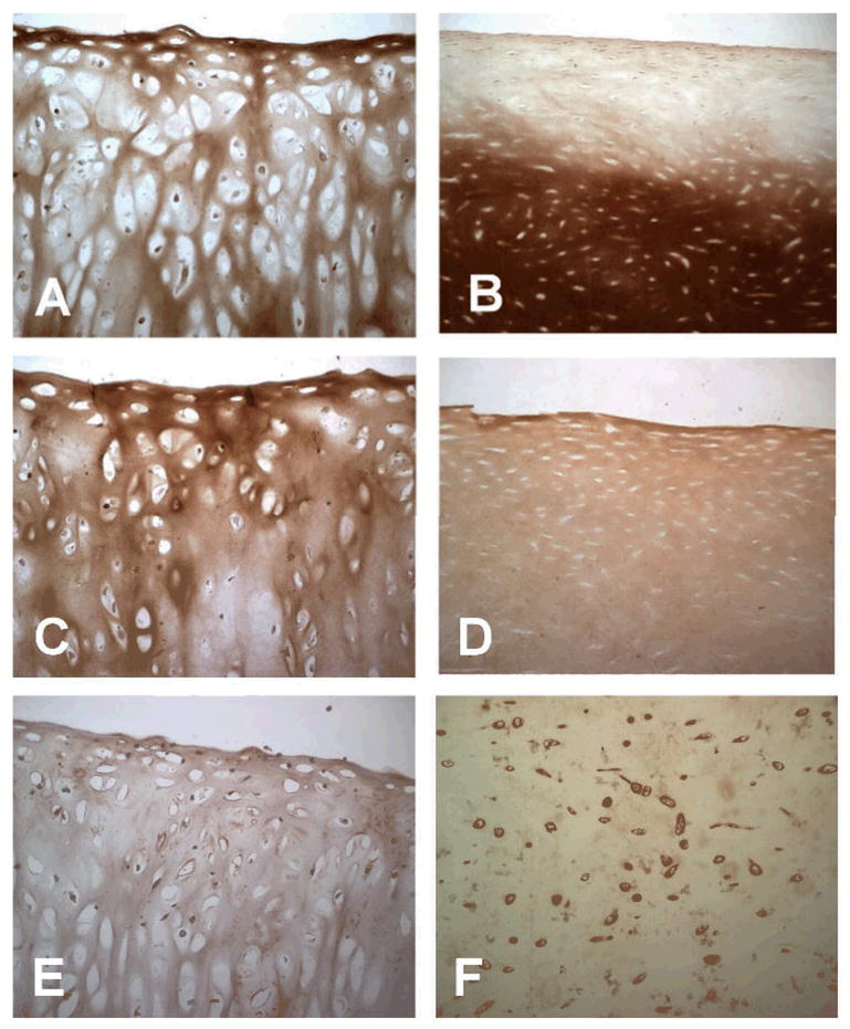
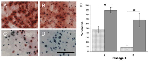
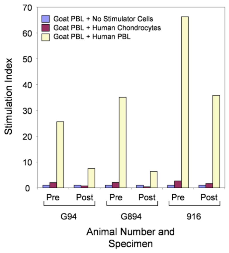
Results: Proteoglycan content in neocartilage produced by juvenile chondrocytes was 100-fold higher than in neocartilage produced by adult cells. Collagen type II and type IX mRNA in fresh juvenile chondrocytes were 100- and 700-fold higher, respectively, than in adult chondrocytes. The distributions of collagens II and IX were similar in native juvenile cartilage and in neocartilage made by juvenile cells. Juvenile cells grew significantly faster in monolayer cultures than adult cells (P = .002) and proteoglycan levels produced in agarose culture was significantly higher in juvenile cells than in adult cells after multiple passages (P < .001). Juvenile chondrocytes did not stimulate lymphocyte proliferation.

Conclusion: These results document a dramatic age-related decline in human chondrocyte chondrogenic potential and show that allogeneic juvenile chondrocytes do not stimulate an immunologic response in vivo.

Clinical relevance: Juvenile human chondrocytes have greater potential to restore articular cartilage than adult cells, and may be transplanted without the fear of rejection, suggesting a new allogeneic approach to restoring articular cartilage in older individuals.


Figures

Figure 1
Figure 1
Chondrocytes from juvenile human articular cartilage (<10 years) show significantly greater matrix synthesis potential than chondrocytes isolated from skeletally mature individuals. Neocartilage was grown under defined serum-free conditions as described previously. Matrix proteoglycan content of newly synthesized neocartilage was measured colorimetrically using dimethylmethylene blue after papain digestion of day 44 to 60 grafts. Data are expressed as the average daily amount of proteoglycan deposited in the tissue matrix after normalization to DNA content. The rate of total sulfated glycosaminoglycan (S-GAG) synthesis for juvenile chondrocytes was more than 100-fold greater than that of adult chondrocytes. For reasons that remain unclear, this potent biologic activity appears to decrease with age and may be lost at pubescence.
Figure 2
Figure 2
Real-time polymerase chain reaction analysis demonstrates 100-fold and 700-fold greater amounts of mRNA for type II (A) and type IX (B) collagen, respectively, in freshly isolated chondrocytes derived from juvenile versus adult human articular cartilage. Gene expression profile analysis of chondrocyte-specific genes was performed as described in the Methods section using glyceraldehyde-3-phosphate dehydrogenase (GAPDH) for data normalization. A marked reduction in collagen gene expression of adult chondrocytes was observed independent of SOX9 expression (C), the master chondrogenic transcription factor of cartilage, which was expressed at approximately the same order of magnitude in juvenile and adult chondrocytes (approximately 83% greater in adult). The ratio of COL2A1/SOX9 mRNA (D) was reduced 200-fold in adult chondrocytes as compared with juvenile articular chondrocytes (n = 5 for juvenile chondrocytes, mean age = 5.6 months, range = 1 day-19 months; n = 3 for adult chondrocytes, mean age = 50.7 years, range = 35–70 years].
Figure 3
Figure 3
Immunohistochemical staining of neocartilage collagen. Formalin-fixed paraffin-embedded sections of human neocartilage (A, C, and E) and native human articular cartilage (B, D, and F) were stained as described in the Methods section for collagen type II (A and B), collagen type IX (C and D), and collagen type VI (E and F). Uniform staining of neocartilage extracellular matrix was observed for collagen types II and IX, whereas weaker staining for type VI collagen demonstrated pericellular localization, similar to what was observed for native articular cartilage obtained from a 10-year-old individual. Original magnification 100×.
Figure 4
Figure 4
Chondrocyte growth rate. A, growth rate (population doublings [PDs] per day) was calculated using cell count data from serially passaged monolayer cultures of chondrocytes from donors of various ages (<1 year old to 78 years old, mean = 34 years). B, the columns show the mean growth rates for chondrocytes derived from juvenile (n = 5) and adult donors (n = 9). Error bars indicate standard deviations. The growth rate for juvenile cells was significantly greater than adult cells (P = .002).
Figure 5
Figure 5
Extracellular matrix accumulation in agarose cultures. Representative areas of sections of agarose-cultured cells derived from juvenile (A and B) or adult donors (C and D) are shown after 2 passages or 3 to 5 population doublings (A and C) or 3 passages or 5 or 6 population doublings (B and D). The black bar in D is 125 μm in length. Percentage of safranin O positive cells were counted microscopically as described in the Methods section for cultures of adult chondrocytes (stippled columns) and juvenile chondrocytes (solid gray columns) at the indicated passages (E). Lines and asterisks above the columns indicate statistically significant differences (P < .001). Error bars represent standard deviations.
Figure 6
Figure 6
Effect of population growth on proteoglycan production. Total sulfated glycosaminoglycan (S-GAG) content was measured in agarose cultures initiated after up to 35 population doublings in monolayer culture. Data are shown for 3 juvenile cell populations and 4 adult cell populations. Error bars show standard deviations of replicate S-GAG assay measurements for each sample (n = 6).
Figure 7
Figure 7
Mixed lymphocyte/chondrocyte reaction (MLCR) culture analysis for potential priming of host lymphocytes after human neocartilage transplantation demonstrates a lack of sensitization 12 weeks after implantation. Cell-based immunity against the implanted grafts was screened 12 weeks after implantation using the MLCR. Goat lymphocytes collected before and after implantation were used as responder cells for analysis as described in the Methods section. No evidence of lymphocyte priming by the implanted neocartilage-derived chondrocytes was observed, despite the fact that goat lymphocytes displayed potent proliferative responses to human peripheral blood leukocytes (PBLs) used as the positive control (ranging from 7-fold to 65-fold above background).

References

    1. Adkisson HD, Gillis MP, Davis EC, Maloney W, Hruska KA. In vitro generation of scaffold independent neocartilage. Clin Orthop Relat Res. 2001 Oct;(391 Suppl):S280–294. - PubMed
    1. Adkisson HD, Milliman C, Zhang X, Mauch K, Maziarz RT, Streeter PR. Immune evasion by neocartilage derived articular chondrocytes. Stem Cell Res. 2009 Accepted. - PubMed
    1. Aigner T, Gebhard PM, Schmid E, Bau B, Harley V, Poschl E. SOX9 expression does not correlate with type II collagen expression in adult articular chondrocytes. Matrix Biol. 2003 Jun;22(4):363–372. - PubMed
    1. Aston JE, Bentley G. Repair of articular surfaces by allografts of articular and growth-plate cartilage. J Bone Joint Surg Br. 1986 Jan;68(1):29–35. - PubMed
    1. Budde B, Blumbach K, Ylostalo J, et al. Altered integration of matrilin-3 into cartilage extracellular matrix in the absence of collagen IX. Mol Cell Biol. 2005 Dec;25(23):10465–10478. - PMC - PubMed

Publication types

MeSH terms