. 2010 May;120(5):1617-26.
doi: 10.1172/JCI41678. Epub 2010 Apr 26.

Heat generates oxidized linoleic acid metabolites that activate TRPV1 and produce pain in rodents

Affiliations

Heat generates oxidized linoleic acid metabolites that activate TRPV1 and produce pain in rodents

Amol M Patwardhan et al. J Clin Invest. 2010 May.

Abstract

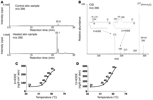
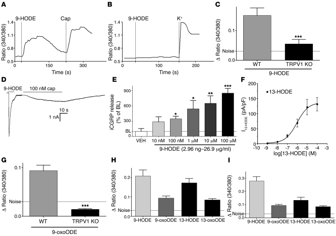
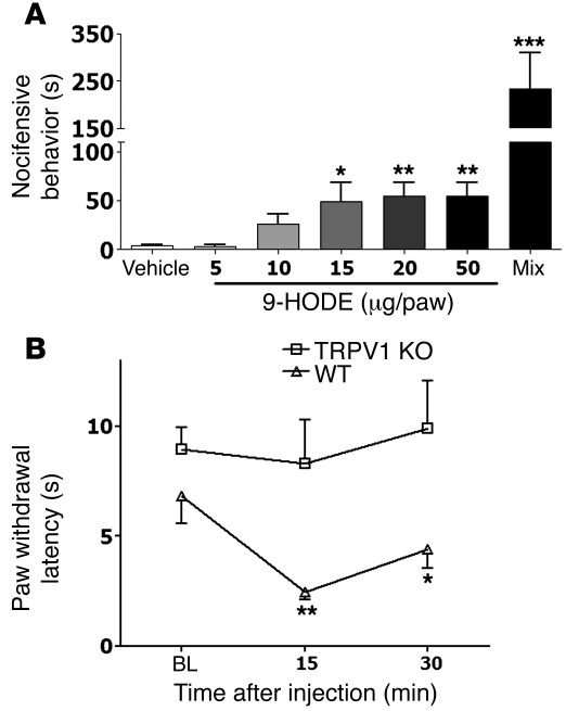
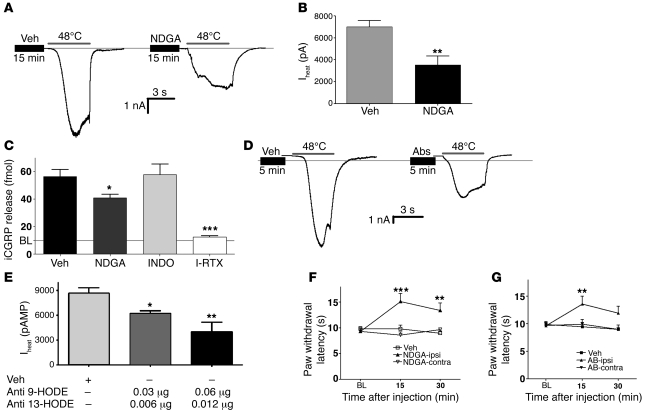
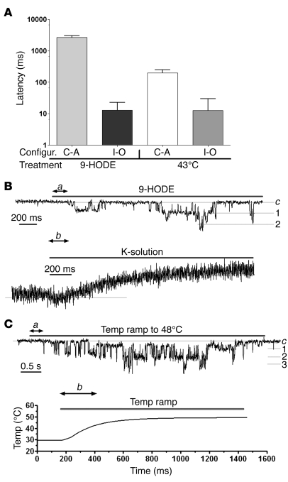
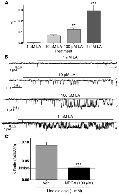
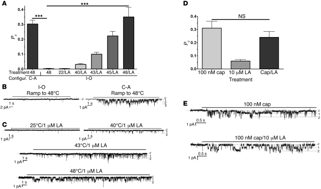
The transient receptor potential vanilloid 1 (TRPV1) channel is the principal detector of noxious heat in the peripheral nervous system. TRPV1 is expressed in many nociceptors and is involved in heat-induced hyperalgesia and thermoregulation. The precise mechanism or mechanisms mediating the thermal sensitivity of TRPV1 are unknown. Here, we have shown that the oxidized linoleic acid metabolites 9- and 13-hydroxyoctadecadienoic acid (9- and 13-HODE) are formed in mouse and rat skin biopsies by exposure to noxious heat. 9- and 13-HODE and their metabolites, 9- and 13-oxoODE, activated TRPV1 and therefore constitute a family of endogenous TRPV1 agonists. Moreover, blocking these substances substantially decreased the heat sensitivity of TRPV1 in rats and mice and reduced nociception. Collectively, our results indicate that HODEs contribute to the heat sensitivity of TRPV1 in rodents. Because oxidized linoleic acid metabolites are released during cell injury, these findings suggest a mechanism for integrating the hyperalgesic and proinflammatory roles of TRPV1 and linoleic acid metabolites and may provide the foundation for investigating new classes of analgesic drugs.


Figures

Figure 1
Figure 1. Heated skin evokes endogenous TRPV1 ligand(s).
(A) Effect of superfusates, collected from 6 mouse skin biopsies (1.5 × 1.5 cm) after exposure to noxious (48°C for 20 min) or control temperatures (37°C for 20 minutes), to TG neurons from WT mice (positive control: capsaicin [Cap], 100 nM) using single-cell calcium imaging. Superfusates were applied after cooling to room temperature. Results are plotted as the 340/380 ratio. (B) The effect of same superfusate (1A) on calcium levels in TG neurons from TRPV1 KO mice. Positive control: MO (50 μM). (C) Graph summarizing comparison of heated skin superfusate application to WT or TRPV1 KO neurons (n = 48 for WT and 36 for TRPV1 KO; P = 0.00001). Mean ± SEM. Results are plotted as Δ ratio 340/380 (Δ = maximum peak 340/380 – baseline 340/380). (D) Temperature dependence of endogenous TRPV1 agonist(s) release (no. of responder/total no. of neurons). (E) Effect of I-RTX (200 nM) pretreatment on superfusate-evoked (mouse skin, heated at 48°C for 20 minutes) responses in TG neurons from WT mice (n = 44 for vehicle [Veh], 31 for I-RTX; P = 0.0003). (F) Heated skin superfusate applied to TRPV1 CHO cells. Positive control: capsaicin (100 nM). (G) Graph summarizing comparison of heated skin superfusate effect on CHO cells expressing TRPV1 (n = 56 for TRPV1, 43 for GFP, negative control; P = 0.00001). (H) Representative inward current in rat TG neuron by heated mouse skin superfusate and capsaicin. (I) Nocifensive behavior (WT vs. TRPV1 KO mice) evoked by hind paw injection of compound(s) isolated from previously heated skin biopsies (n = 5–6 per group; P = 0.0003). ***P < 0.001.
Figure 2
Figure 2. Heating increases oxidized linoleic acid metabolites in skin.
(A) HPLC comparison of unique substances in superfusates collected from mouse skin biopsies (1.5 × 1.5 cm) after exposure to noxious heat (48°C for 20 min) or control temperatures (37°C for 20 min). The TRPV1 activity of each fraction was evaluated using calcium imaging of TG neurons cultured from rats and CHO cells expressing TRPV1 (data not shown). cps, counts per second. (B) Evaluation of product ions formed from HPLC fraction 22 following collisional activation of the [M-H]_ ion at m/z 295 with tandem quadrupole mass spectrometer monitoring of either m/z 171 (9-HODE) or m/z 195 (13-HODE). (C and D) Temperature-dependent release of 9-HODE (C) and 13-HODE (D) into mouse skin superfusates collected after 20 minutes exposure to a given temperature. 9- and 13-HODE were detected by HPLC/MS as described (x axis has a logarithmic scale).
Figure 3
Figure 3. Oxidized linoleic acid metabolites are TRPV1 agonists.
(A and B) Synthetic 9-HODE (100 μM) activates TG neurons from WT mice but not from TRPV1 KO mice as measured using calcium imaging. (C) Graph summarizing comparison of 9-HODE activation of TG neurons from WT versus TRPV1 KO mice (n = 50 for WT and 66 for TRPV1 KO; P = 0.0002). (D) Whole-cell recording demonstrates activation of a rat TG neuron by synthetic 9-HODE (100 μM) and capsaicin. (E) Concentration-response effects of applying synthetic 9-HODE (15 minutes) on iCGRP release from cultured rat TG neurons as measured by radioimmunoassay (n = 8–16 wells/group; P = 0.0001). BL, baseline. (F) Concentration-response curve for synthetic 13-HODE. Responses (pA/pF) were recorded from TRPV1-expressing CHO cells. (G) Effect of applying synthetic 9-oxoODE (100 μM) on TG neurons from WT mice (n = 71 for WT and 83 for TRPV1 KO; P = 0.0007). *P < 0.05; **P < 0.01; ***P < 0.001. (H and I) Comparison of the effects of applying linoleic acid metabolites (all 100 μM; n = 47–75) to TG neurons cultured from WT mice (H) or to CHO cells transfected with TRPV1 (I) as measured by calcium imaging.
Figure 4
Figure 4. Oxidized linoleic acid metabolites trigger acute pain and thermal hyperalgesia.
(A) Effects of an ipl hind paw injection in rats of 9-HODE or a mixture of 9-HODE, 13-HODE, 9-oxoODE, and 13-oxoODE (mix, 25 μg each) on spontaneous nocifensive behavior (duration of flinches; n = 4–6/group; *P < 0.05; **P = 0.003 vs. vehicle; ***P = 0.0001 vs. all other groups). Observers were blinded to treatment allocation. (B) Effects of an ipl injection of 9-HODE in WT or TRPV KO mice on paw withdrawal latency to a beam of radiant heat (n = 6 per group; P = 0.007 at 15 minutes).
Figure 5
Figure 5. Inhibition of oxidized linoleic acid metabolites decreases heat sensitivity of TRPV1.
(A) Effect of applying vehicle or NDGA (30 μM, 15 minutes) on whole-cell heat-evoked inward current (Iheat) in a cultured rat TG neuron. (B) Summary graph shows the effects of NDGA on Iheat in rat TG neurons (n = 19 for vehicle, 9 for NDGA; P = 0.0018). (C) The effect of pre- and cotreatment with NDGA (10 μM), indomethacin (INDO) (2 μM), and I-RTX (200 nM) on heat-evoked iCGRP release from cultured rat TG neurons (n = 8–16 wells/group; P = 0.02 for NDGA and 0.00001 for I-RTX). (D) Effect of an intracellular dialysis (for 5–10 minutes) of either vehicle or a combination of anti–9-HODE and anti–13-HODE antibodies (0.06 and 0.012 μg, respectively) on Iheat in cultured rat TG neurons. (E) Graph summarizing effects of dialysis with antibodies against 9- and 13-HODE on Iheat in cultured rat TG neurons (n = 15 for vehicle and 7 for each antibody mix group, P = 0.007; antibody dose refers to amount in a recording pipette). (F and G) Effect of ipl hind paw injection of vehicle, NDGA (F, n = 6/group; P = 0001), or a mixture of anti–9-HODE and anti–13-HODE antibodies (G, 25 μg each/paw, n = 6/group; P = 0.005) on paw withdrawal latencies to a beam of radiant heat in rats. Observers were blinded to treatment allocation. *P < 0.05; **P < 0.01; ***P < 0.001.
Figure 6
Figure 6. Application of 9-HODE and heat (43°C) demonstrate similar latencies for activation of TRPV1.
(A) The latencies for generation of single-channel inward currents due to application of either 9-HODE (100 μM) or 43°C to TRPV1-expressing CHO cells. n = 6–10. Recording was carried out in cell-attached (C-A) and inside-out (I-O) configurations (configur.). (B) Representative single-channel inside-out traces illustrating the measurement of the latency for 9-HODE. Latency was calculated as the difference between a and b. A solution containing potassium (50 mM) was applied via the recording pipette to measure the inherent latency of the perfusion system (b). Inside-out configuration was established for 3–5 minutes. c is a close state. 1, 2, and 3 are numbers of channels in the patches. (C) Representative single-channel inside-out traces illustrating the measurement of the latency for 43°C. Temperature was applied immediately (within 1 minute) after establishment of the inside-out configuration. Latency was calculated as ab, where b is the time to reach 43°C in the vicinity of the cells. Temperature ramp was recorded by placing a thermistor in the position of previously recorded cells.
Figure 7
Figure 7. Indirect activation of TRPV1 by linoleic acid.
(A) Concentration-dependent effect of linoleic acid (LA) on Po of TRPV1 in transfected CHO cells using inside-out configuration at room temperature. Patches were washed at least 3 minutes before linoleic acid application at room temperature. (B) Representative single-channel traces demonstrating the effect of different concentrations of linoleic acid on Po of TRPV1-expressing CHO cell membrane patches. c is a close state. o is an open state, in which the patch contained 1 channel. 1, 2, and 3 are the numbers of channels in the patches. (C) Effect of pretreatment with either vehicle or NDGA (30 μM, 15 minutes) on linoleic acid–induced activation of TRPV1 transfected CHO cells as measured using calcium imaging (n = 25–50; P = 0.0001). **P < 0.01; ***P < 0.001.
Figure 8
Figure 8. Linoleic acid rescues the TRPV1-mediated heat responses in membrane patches.
(A) The responsiveness of TRPV1-expressing CHO cells to 48°C is maintained in the cell-attached configuration but is lost by 3 minutes of washing the inside-out configuration. The application of heated linoleic acid (LA) (1 μM) rescues temperature responses (i.e., Po) in inside-out patches from TRPV1-expressing cells that were washed at least 3 minutes. Single-channel configurations and applied temperatures are indicated. (n = 4–8; P = 0.0001). ***P < 0.001. (B) Representative traces show single-channel recording at indicated temperatures in inside-out and cell-attached configurations. Inside-out patches were washed at least 3 minutes prior to the recording. (C) Representative traces illustrate the effects of application of 1 μM LA at indicated temperatures. Recordings were performed from inside-out patches washed at least 3 minutes. (D) Coapplication of 10 μM linoleic acid does not affect capsaicin-gated (100 nM) responses in TRPV1-expressing patches. Recordings were performed from inside-out patches washed at least 3 minutes. (E) Representative traces show single-channel recording for Cap and a mix of Cap and linoleic acid.

Comment in

References

    1. Brauchi S, Orta G, Salazar M, Rosenmann E, Latorre R. A hot-sensing cold receptor: C-terminal domain determines thermosensation in transient receptor potential channels. J Neurosci. 2006;26(18):4835–4840. doi: 10.1523/JNEUROSCI.5080-05.2006. - DOI - PMC - PubMed
    1. Caterina MJ, Schumacher MA, Tominaga M, Rosen TA, Levine JD, Julius D. The capsaicin receptor: a heat-activated ion channel in the pain pathway. Nature. 1997;389(6653):816–824. doi: 10.1038/39807. - DOI - PubMed
    1. Lumpkin EA, Caterina MJ. Mechanisms of sensory transduction in the skin. Nature. 2007;445(7130):858–865. doi: 10.1038/nature05662. - DOI - PubMed
    1. Tominaga M, et al. The cloned capsaicin receptor integrates multiple pain-producing stimuli. Neuron. 1998;21(3):531–543. doi: 10.1016/S0896-6273(00)80564-4. - DOI - PubMed
    1. Voets T, Droogmans G, Wissenbach U, Janssens A, Flockerzi V, Nilius B. The principle of temperature-dependent gating in cold- and heat-sensitive TRP channels. Nature. 2004;430(7001):748–754. doi: 10.1038/nature02732. - DOI - PubMed

Publication types