. 2010 May;120(5):1570-7.
doi: 10.1172/JCI40267. Epub 2010 Apr 26.

A regulatory feedback loop involving p63 and IRF6 links the pathogenesis of 2 genetically different human ectodermal dysplasias

Affiliations

A regulatory feedback loop involving p63 and IRF6 links the pathogenesis of 2 genetically different human ectodermal dysplasias

Francesca Moretti et al. J Clin Invest. 2010 May.

Abstract

The human congenital syndromes ectrodactyly ectodermal dysplasia-cleft lip/palate syndrome, ankyloblepharon ectodermal dysplasia clefting, and split-hand/foot malformation are all characterized by ectodermal dysplasia, limb malformations, and cleft lip/palate. These phenotypic features are a result of an imbalance between the proliferation and differentiation of precursor cells during development of ectoderm-derived structures. Mutations in the p63 and interferon regulatory factor 6 (IRF6) genes have been found in human patients with these syndromes, consistent with phenotypes. Here, we used human and mouse primary keratinocytes and mouse models to investigate the role of p63 and IRF6 in proliferation and differentiation. We report that the DeltaNp63 isoform of p63 activated transcription of IRF6, and this, in turn, induced proteasome-mediated DeltaNp63 degradation. This feedback regulatory loop allowed keratinocytes to exit the cell cycle, thereby limiting their ability to proliferate. Importantly, mutations in either p63 or IRF6 resulted in disruption of this regulatory loop: p63 mutations causing ectodermal dysplasias were unable to activate IRF6 transcription, and mice with mutated or null p63 showed reduced Irf6 expression in their palate and ectoderm. These results identify what we believe to be a novel mechanism that regulates the proliferation-differentiation balance of keratinocytes essential for palate fusion and skin differentiation and links the pathogenesis of 2 genetically different groups of ectodermal dysplasia syndromes into a common molecular pathway.


Figures

Figure 1
Figure 1. Irf6 is a direct p63 target.
(A) Microarray analysis of Irf6 RNA expression in mouse primary keratinocytes transfected with ΔNp63- or TAp63-specific siRNAs. Representative heat map and raw value data are shown. (B) Irf6 mRNA expression in mouse primary keratinocytes transfected with ΔNp63- or TAp63-specific siRNA. Data are presented as mean ± SEM. *P = 0.001. (C) p63 REs present within the Irf6 gene. The p63 RE consensus sequence is shown at the bottom. Arrowheads indicate the position of primers used in ChIP analysis. Putative p53/p63 REs were identified by PathSearch algorithm. (D) ChIP analysis to detect p63 occupying the Irf6 promoter in differentiating mouse primary keratinocytes. PCR was performed using the indicated primers (Supplemental Methods). (E) IRF6 expression in human primary keratinocytes by RT-qPCR. Data are presented as mean ± SEM. (F) Immunoblot analysis of human primary keratinocyte protein extracts to detect IRF6, ΔNp63, K1, and PCNA proteins. (G) IRF6 expression in human primary keratinocytes transfected with the indicated expression vectors and induced to differentiate. ΔNp63α induces IRF6 mRNA. Data are presented as mean ± SEM. cons., consensus sequence; ex., exon; mIrf6, mouse Irf6; siCtr., control siRNA; siΔNp63, ΔNp63-specific siRNA; siTAp63, TAp63-specific siRNA.
Figure 2
Figure 2. p63 is required for Irf6 expression in vivo.
(A) The palatal phenotype in E13.5 and newborn p63–/– embryos. The age is indicated at the top columns, and genotypes are reported to the left of rows. The images on the far right of each row illustrate the newborn specimens, after removal of the mandible (Md). The dashed black lines define the profile of the anatomical structures. In p63–/– embryos, the maxillae (Mx) and the secondary palate (pal II) fail to fuse with the primary palate (pal I) and the secondary palate fails to fuse on the midline (thin dashed red line, red asterisks). In situ hybridization for Msx1 and Irf6 on coronal sections of the tooth (E13.5, left) and palatal (E13.5, right) region of wild-type and p63–/– embryos. The section plane is indicated by thick dashed red lines. Expression of Msx1 is indicated with red arrows. Expression of Irf6 is reduced in the tooth and palate epithelia of the p63–/– specimen (black arrows and asterisks for wild-type and mutant, respectively). Scale bars: 1 mm (white); 50 μm (black). T, tongue. (B) RT-qPCR analysis of Irf6 mRNA in ectoderm of p63–/– mice aged E13.5 and EEC mice aged E16.5. The wild-type value is set as 1. Data are presented as mean ± SEM. *P = 0.01. (C) Skin sections from E16.5 wild-type (+/+) or p63+/EEC knock-in mice, immunostained with Irf6- or p63-specific antibodies. Irf6 immunostaining is strongly reduced in p63+/EEC mice, while p63 expression is increased. Images are representative of data obtained from 3 littermates for each genotype. Scale bars: 30 μm.
Figure 3
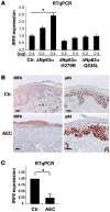
Figure 3. p63 mutations affect IRF6 expression in vitro and in vivo.
(A) IRF6 expression in H1299 cells transfected with ΔNp63α (WT), ΔNp63α H279R (EEC), and ΔNp63α Q536L (AEC) expression vectors. Data are presented as mean ± SEM. *P = 0.01. (B) Skin sections from a newborn affected by AEC syndrome or from normal subjects (Ctr), immunostained with IRF6- and p63-specific antibodies. IRF6 expression is strongly downregulated in AEC epidermis. Images are representative of data obtained from 3 unrelated normal subjects and from 3 independent sections from the AEC patient. Scale bars: 30 μm. (C) RT-qPCR showing IRF6 mRNA downregulation in total RNA extracted from formalin-fixed, paraffin-embedded sections. Results were normalized against hARP. Data are presented as mean ± SEM. *P = 0.001.
Figure 4
Figure 4. IRF6 induces downregulation of ΔNp63 in human primary keratinocytes.
(A) Dual immunofluorescence on human primary keratinocytes transfected with a HA-tag–IRF6 expression vector, with anti-HA and anti-p63 antibodies, to detect double-positive cells. Exogenous IRF6 decreased endogenous p63 expression. Arrowheads indicate transfected cells. Cell counts are reported in Table 1. Scale bar: 20 μm. (B) IRF6-depleted keratinocytes fail to downregulate ΔNp63 expression. Human primary keratinocytes were transfected with control (siCtr) or IRF6-specific (siIRF6) siRNA, followed by immunoblot detection of ΔNp63, IRF6, and PCNA. ΔNp63 was quantified by the ImageJ software, normalized, and expressed as fold induction (FI). (C) Immunoblot detection of HA-tag–IRF6 and ΔNp63α in U2OS cells transfected with HA-IRF6 or ΔNp63α expression plasmids and treated with the proteasome inhibitor MG132 (5 μM, 6 hours). MG132 treatment inhibited ΔNp63 downregulation by IRF6. (D) Immunoblot detection of ΔNp63α and IRF6 in H1299 cells transfected with a vector expressing either wild-type ΔNp63α or mutated ΔNp63α-R279H, in the presence or absence of HA-IRF6. ΔNp63α-R279H is resistant to IRF6-mediated downregulation. (E) Immunoblot detection of ΔNp63α and IRF6 in H1299 cells transfected with a ΔNp63α expression vector, in the presence of either wild-type or R84C mutant IRF6. The IRF6-R84C mutant failed to induce ΔNp63α downregulation. Results are representative of 3 independent experiments.
Figure 5
Figure 5. IRF6 negatively modulates the proliferative potential of epithelial cells.
(A) Colony formation of TE13 epithelial carcinoma cells transfected with the indicated expression vectors (ΔNp63α, ΔNp63α/R279H, or HA-IRF6). Colonies of more than 1 mm in diameter were counted, and results are expressed as mean ± SEM (3 independent experiments, duplicate samples). IRF6 expression induced growth arrest that is partially reverted by ΔNp63α-R279H coexpression. (B) Colony formation of human primary keratinocytes transduced with a vector expressing HA-tag–IRF6. Immunoblotting shows HA-IRF6 expression. Actin was used as loading control. The number of colonies that were more than 1 mm in diameter was calculated with ImageJ software. IRF6 expression reduced the proliferative potential of keratinocytes. Results are expressed as percentage of each kind of colony over the total (error bars = 1 SD). P = 0.01, control versus IRF6 (3 independent experiments, duplicate samples). The relative percentage of holoclones, paraclones, and meroclones is shown in Table 2. (C) Schematic representation of the link between ΔNp63 and IRF6. IRF6 is transcriptionally activated by ΔNp63 during differentiation. IRF6 protein expression induces ΔNp63 degradation, reducing proliferative potential of epithelial cells.

Comment in

References

    1. Yang A, et al. p63, a p53 homolog at 3q27-29, encodes multiple products with transactivating, death-inducing, and dominant-negative activities. Mol Cell. 1998;2(3):305–316. doi: 10.1016/S1097-2765(00)80275-0. - DOI - PubMed
    1. Yang A, et al. p63 is essential for regenerative proliferation in limb, craniofacial and epithelial development. Nature. 1999;398(6729):714–718. doi: 10.1038/19539. - DOI - PubMed
    1. Ghioni P, Bolognese F, Duijf PH, Van Bokhoven H, Mantovani R, Guerrini L. Complex transcriptional effects of p63 isoforms: identification of novel activation and repression domains. Mol Cell Biol. 2002;22(24):8659–8668. doi: 10.1128/MCB.22.24.8659-8668.2002. - DOI - PMC - PubMed
    1. Dohn M, Zhang S, Chen X. p63alpha and DeltaNp63alpha can induce cell cycle arrest and apoptosis and differentially regulate p53 target genes. Oncogene. 2001;20(25):3193–3205. doi: 10.1038/sj.onc.1204427. - DOI - PubMed
    1. Koster MI, Kim S, Roop DR. p63 deficiency: a failure of lineage commitment or stem cell maintenance? . J Investig Dermatol Symp Proc. 2005;10(2):118–123. doi: 10.1111/j.1087-0024.2005.200416.x. - DOI - PubMed

Publication types