. 2010 May 25;102(11):1636-44.
doi: 10.1038/sj.bjc.6605668. Epub 2010 Apr 27.

Molecular characterisation of side population cells with cancer stem cell-like characteristics in small-cell lung cancer

Affiliations

Molecular characterisation of side population cells with cancer stem cell-like characteristics in small-cell lung cancer

C D Salcido et al. Br J Cancer. .

Abstract

Background: Side population (SP) fraction cells, identified by efflux of Hoechst dye, are present in virtually all normal and malignant tissues. The relationship between SP cells, drug resistance and cancer stem cells is poorly understood. Small-cell lung cancer (SCLC) is a highly aggressive human tumour with a 5-year survival rate of <10%. These features suggest enrichment in cancer stem cells.

Methods and results: We examined several SCLC cell lines and found that they contain a consistent SP fraction that comprises <1% of the bulk population. Side population cells have higher proliferative capacity in vitro, efficient self-renewal and reduced cell surface expression of neuronal differentiation markers, CD56 and CD90, as compared with non-SP cells. Previous reports indicated that several thousand SP cells from non-small-cell lung cancer are required to form tumours in mice. In contrast, as few as 50 SP cells from H146 and H526 SCLC cell lines rapidly reconstituted tumours. Whereas non-SP cells formed fewer and slower-growing tumours, SP cells over-expressed many genes associated with cancer stem cell and drug resistance: ABCG2, FGF1, IGF1, MYC, SOX1/2, WNT1, as well as genes involved in angiogenesis, Notch and Hedgehog pathways.

Conclusions: Side population cells from SCLC are highly enriched in tumourigenic cells and are characterised by a specific stem cell-associated gene expression signature. This gene signature may be used for development of targeted therapies for this rapidly fatal tumour.


Figures
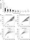
Figure 1
Figure 1
Characterisation of cells from SP and non-SP fractions. (A) Side population cells have higher proliferative capacity in vitro. H146 cells were sorted from SP and non-SP populations and plated by limiting dilution in six-tuplicate wells. The number of growing colonies at the end of 3 weeks was scored and data (expressed as mean number of colonies per well) were averaged from two independent experiments±s.e. Open bars: SP cells; solid bars: non-SP cells. (B) Cells sorted from SP fraction repopulate the original population. Side population and non-SP cells were sorted according to Verapamil gating (upper panels) and cultured for 17 days (lower panels).
Figure 2
Figure 2
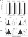
Expression of differentiation markers is decreased in SP fraction. (A) Percent cell surface expression of CD56 and CD90 within the SP and non-SP cells in H146 and H526 cells. Solid bars: SP cells; open bars: non-SP cells±s.d. The differences between each pair are statistically significant as determined by the two-tailed Fisher ’s t-test, indicated by P values above. (B) Cell surface expression in H146 and H526 cells for each marker using PE-labelled antibodies. The dashed line represents cells in SP fraction, and the solid line shows cells in non-SP fraction. The filled area represents unstained cells.
Figure 3
Figure 3
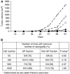
Tumorigenicity of SP and non-SP cells in vivo. (A) In total, 50 (circles), 100 (triangles) and 500 (squares) H146 cells were implanted in triplicate injections and tumour growth rates of SP cells (dashed lines, open symbols) and non-SP cells (solid lines, closed symbols) were monitored biweekly. The lines represent the average tumour volume calculated from one experiment, as indicated in Materials and Methods. (B) Summary of H146 tumours formed following implantation of the indicated cell numbers from SP and non-SP cells from four independent experiments. Small tumours that were only palpable (<5 mm2) were scored as positive.
Figure 4
Figure 4
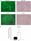
Tumours arising from SP fraction cells a have higher degree of angiogenesis. (A) Microvessel staining using mouse anti-CD31 antibodies of one representative field from tumours generated from 500 non-SP (upper panel) and 500 SP (lower panel) H146 cells. H&E staining for each tumour is showed on the right. (B) Computer-generated quantitative analysis of anti-CD31 staining fluorescence intensity from one representative tumour. Non-SP (filled bars) and SP (open bars). Bars represent s.d. from 9–10 computer-generated random fields from three slides from one representative tumour. The measurements are from one of two individual tumours±s.d.
Figure 5
Figure 5
Gene expression analysis validation by quantitative RT–PCR. Gene expression for SP fraction (open bars) and Non-SP fraction (closed bars) was analysed as described in Materials and Methods. The data are normalised to a human liver as universal standard (±s.d. from triplicate samples) using three sorting experiments representing independent biological replicates. All genes shown were significantly upregulated in SP cells, except for GB1 and JAG1. The graph shows the results in a format (both numerically and graphically) showing expression of each gene in SP and non-SP fractions relative to combination of four housekeeping genes and a reference universal standard (human liver).

References

    1. Al Hajj M, Clarke MF (2004) Self-renewal and solid tumor stem cells. Oncogene 23: 7274–7282 - PubMed
    1. Al Hajj M, Wicha MS, Benito-Hernandez A, Morrison SJ, Clarke MF (2003) Prospective identification of tumorigenic breast cancer cells. Proc Natl Acad Sci USA 100: 3983–3988 - PMC - PubMed
    1. Behbod F, Xian W, Shaw CA, Hilsenbeck SG, Tsimelzon A, Rosen JM (2006) Transcriptional profiling of mammary gland side population cells. Stem Cells 24: 1065–1074 - PubMed
    1. Caussinus E, Gonzalez C (2005) Induction of tumor growth by altered stem-cell asymmetric division in Drosophila melanogaster. Nat Genet 37: 1125–1129 - PubMed
    1. Collins AT, Berry PA, Hyde C, Stower MJ, Maitland NJ (2005) Prospective identification of tumorigenic prostate cancer stem cells. Cancer Res 65: 10946–10951 - PubMed

Publication types

MeSH terms