Review
. 2010 Apr;12(2):156-61.
doi: 10.1007/s11926-010-0095-0.

Imaging lung disease in systemic sclerosis

Affiliations
Review

Imaging lung disease in systemic sclerosis

Diane Strollo et al. Curr Rheumatol Rep. 2010 Apr.

Abstract

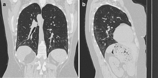
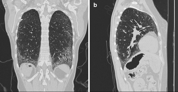
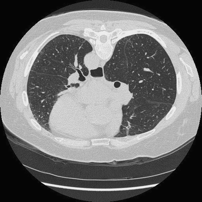
Interstitial lung disease and pulmonary hypertension (PH) are the most common cardiopulmonary findings in patients with systemic sclerosis (SSc). About two thirds of patients suffering from SSc develop scleroderma interstitial lung disease. PH is present in about 20% of SSc patients and is typically associated with severe lung disease, although it may be an isolated manifestation of SSc. High-resolution CT scanning is a key method for evaluating chest involvement. There are four roles of imaging in scleroderma interstitial lung disease: 1) detection of lung involvement, 2) identification of patients likely to respond to treatment, 3) assessment of treatment efficacy, and 4) exclusion of other significant diseases to include PH and cardiac and esophageal abnormalities.


Figures

Fig. 1
Fig. 1
Coronal and sagittal chest CT reconstructions (lung window) demonstrate the classic appearance of early lung involvement in scleroderma interstitial lung disease. Initially subtle alterations of increased ground glass opacity and accentuated reticular markings (a and b) that affect the peripheral, posterior, and basilar portions of the lungs
Fig. 2
Fig. 2
Coronal and sagittal chest CT reconstructions (lung window) demonstrate the classic appearance of scleroderma interstitial lung disease in patients with progressive disease, showing extensive architectural distortion due to progressive pulmonary fibrosis (a and b)
Fig. 3
Fig. 3
Esophageal dilatation with retained debris is an early and frequent manifestation of scleroderma and may help differentiate interstitial lung disease due to connective tissue disease from that of other etiologies

References

    1. Kreig T, Takehara K. Skin disease: a cardinal feature of systemic sclerosis. Rheumatology (Oxford) 2009;48(Suppl 3):iii14–iii18. doi: 10.1093/rheumatology/kep108. - DOI - PubMed
    1. Silver RM, Miller KS. Lung involvement in systemic sclerosis. Rheum Dis Clin North Am. 1990;16:199–216. - PubMed
    1. Ramirez A, Varga J. Pulmonary arterial hypertension in systemic sclerosis: clinical manifestations, pathophysiology, evaluation, and management. Treat Respir Med. 2004;3:339–352. doi: 10.2165/00151829-200403060-00002. - DOI - PubMed
    1. Gilliland BC, et al. Systemic sclerosis (scleroderma) In: Braunwald E, Fauci AS, Kasper DL, et al., editors. Harrison’s Principles of Internal Medicine. 15. New York: McGraw-Hill; 2001.
    1. Arroliga AC, Podell DN, Matthay RA. Pulmonary manifestations of scleroderma. J Thorac Imaging. 1992;7:30–45. doi: 10.1097/00005382-199203000-00005. - DOI - PubMed

Publication types

MeSH terms