Review
. 2010 May;10(3):207-14.
doi: 10.1007/s11910-010-0104-8.

Synapse pathology in psychiatric and neurologic disease

Affiliations
Review

Synapse pathology in psychiatric and neurologic disease

Myrrhe van Spronsen et al. Curr Neurol Neurosci Rep. 2010 May.

Abstract

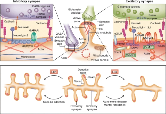
Inhibitory and excitatory synapses play a fundamental role in information processing in the brain. Excitatory synapses usually are situated on dendritic spines, small membrane protrusions that harbor glutamate receptors and postsynaptic density components and help transmit electrical signals. In recent years, it has become evident that spine morphology is intimately linked to synapse function--smaller spines have smaller synapses and support reduced synaptic transmission. The relationship between synaptic signaling, spine shape, and brain function is never more apparent than when the brain becomes dysfunctional. Many psychiatric and neurologic disorders, ranging from mental retardation and autism to Alzheimer's disease and addiction, are accompanied by alterations in spine morphology and synapse number. In this review, we highlight the structure and molecular organization of synapses and discuss functional effects of synapse pathology in brain disease.


Figures

Fig. 1
Fig. 1
Molecular architecture of inhibitory and excitatory synapses. The top panels show excitatory and inhibitory synapses. Excitatory synapses target on mature mushroom-shaped spines containing a prominent postsynaptic density (PSD), and inhibitory synapses are present along the dendritic shaft lacking postsynaptic thickening. Various organelles support the synapse; mitochondria provide energy, polyribosomes and RNA particles allow local protein synthesis, recycling endosomes (REs) transport internalized synaptic receptors back to the plasma membrane, and the cytoskeleton regulates spine dynamics. The abundant actin cytoskeleton is connected to the PSD and is the primary determinant of spine shape and motility. Transient invasion of dynamic microtubule into dendritic spines can regulate formation of spine head protrusions and rapid spine growth. Excitatory and inhibitory synapses contain a unique set of channels, scaffolding proteins, and other postsynaptic molecules. The microanatomy of the inhibitory and excitatory synapses and their organization of proteins and protein–protein interactions are depicted in the left and right panels, respectively. Major families of postsynaptic proteins are shown, including scaffolding proteins, adhesion molecules, and receptors. The lower panel shows major morphologic events occurring in dendritic spines upon long-term potentiation (LTP; left) or long-term depression (LTD; right). In Alzheimer’s disease and mental retardation, signaling cascades are triggered similar to LTD, leading to thinner, immature spines. In contrast, cocaine addiction shows similarities to LTP, resulting in bigger, mushroom-shaped, mature spines. The molecular and morphologic changes in the synapse are hallmarks of the disease pathology and are responsible for the cognitive alterations in neuropsychiatric diseases. CamKII—Ca2+/calmodulin-dependent kinase II; AMPAR—amino-3-hydroxy-5-methyl-4-isoazolepropionate receptor; GABA—γ-aminobutyric acid; GABAR—GABA receptor; NMDARN-methyl-d-aspartate receptor; mGluR—metabotropic glutamate receptor; SAPAP—synapse-associated protein 90/PSD-95-associated protein

References

    1. Yuste R, Bonhoeffer T. Morphological changes in dendritic spines associated with long-term synaptic plasticity. Annu Rev Neurosci. 2001;24:1071–1089. doi: 10.1146/annurev.neuro.24.1.1071. - DOI - PubMed
    1. Pfeiffer BE, Huber KM. The state of synapses in fragile X syndrome. Neuroscientist. 2009;15(5):549–567. doi: 10.1177/1073858409333075. - DOI - PMC - PubMed
    1. Stephan KE, Baldeweg T, Friston KJ. Synaptic plasticity and dysconnection in schizophrenia. Biol Psychiatry. 2006;59(10):929–939. doi: 10.1016/j.biopsych.2005.10.005. - DOI - PubMed
    1. Calabresi P, Picconi B, Parnetti L, Di Filippo M. A convergent model for cognitive dysfunctions in Parkinson’s disease: the critical dopamine-acetylcholine synaptic balance. Lancet Neurol. 2006;5(11):974–983. doi: 10.1016/S1474-4422(06)70600-7. - DOI - PubMed
    1. Sudhof TC. Neuroligins and neurexins link synaptic function to cognitive disease. Nature. 2008;455(7215):903–911. doi: 10.1038/nature07456. - DOI - PMC - PubMed

Publication types