

Mechanisms involved in injury and repair of the murine lacrimal gland: role of programmed cell death and mesenchymal stem cells
- PMID: 20427009
- PMCID: PMC3225027
- DOI: 10.1016/s1542-0124(12)70070-8
Mechanisms involved in injury and repair of the murine lacrimal gland: role of programmed cell death and mesenchymal stem cells
Erratum in
- Ocul Surf. 2010 Jul;8(3):134
Abstract
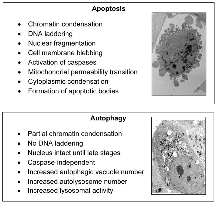
The non-keratinized epithelia of the ocular surface are constantly challenged by environmental insults, such as smoke, dust, and airborne pathogens. Tears are the sole physical protective barrier for the ocular surface. Production of tears in inadequate quantity or of inadequate quality results in constant irritation of the ocular surface, leading to dry eye disease, also referred to as keratoconjunctivitis sicca (KCS). Inflammation of the lacrimal gland, such as occurs in Sjogren syndrome, sarcoidosis, chronic graft-versus-host disease, and other pathological conditions, results in inadequate secretion of the aqueous layer of the tear film and is a leading cause of dry eye disease. The hallmarks of lacrimal gland inflammation are the presence of immune cell infiltrates, loss of acinar epithelial cells (the secreting cells), and increased production of proinflammatory cytokines. To date, the mechanisms leading to acinar cell loss and the associated decline in lacrimal gland secretion are still poorly understood. It is also not understood why the remaining lacrimal gland cells are unable to proliferate in order to regenerate a functioning lacrimal gland. This article reviews recent advances in exocrine tissue injury and repair, with emphasis on the roles of programmed cell death and stem/progenitor cells.
Figures


References
-
- Holly FJ. Tear film physiology. Int Ophthalmol Clin. 1987;27:2–6. - PubMed
-
- Tiffany JM. The normal tear film. Dev Ophthalmol. 2008;41:1–20. - PubMed
-
- Mircheff AK. Water and electrolyte secretion and fluid modification. In: Albert D, Jakobiec F, editors. Principles and practice of ophthalmology: basic sciences. Philadelphia: WB Saunders; 1994. pp. 466–72.
-
- Lemullois M, Rossignol B, Mauduit P. Immunolocalization of myoepithelial cells in isolated acini of rat exorbital lacrimal gland: cellular distribution of muscarinic receptors. Biol Cell. 1996;86:175–81. - PubMed
Publication types
MeSH terms
Grants and funding
LinkOut - more resources
Full Text Sources
Other Literature Sources
