. 2010 Jun 25;285(26):20109-16.
doi: 10.1074/jbc.M110.121699. Epub 2010 Apr 28.

Tti1 and Tel2 are critical factors in mammalian target of rapamycin complex assembly

Affiliations

Tti1 and Tel2 are critical factors in mammalian target of rapamycin complex assembly

Takeshi Kaizuka et al. J Biol Chem. .

Abstract

Mammalian target of rapamycin (mTOR) is a member of the phosphatidylinositol 3-kinase-related kinase (PIKK) family and is a major regulator of translation, cell growth, and autophagy. mTOR exists in two distinct complexes, mTORC1 and mTORC2, that differ in their subunit composition. In this study, we identified KIAA0406 as a novel mTOR-interacting protein. Because it has sequence homology with Schizosaccharomyces pombe Tti1, we named it mammalian Tti1. Tti1 constitutively interacts with mTOR in both mTORC1 and mTORC2. Knockdown of Tti1 suppresses phosphorylation of both mTORC1 substrates (S6K1 and 4E-BP1) and an mTORC2 substrate (Akt) and also induces autophagy. S. pombe Tti1 binds to Tel2, a protein whose mammalian homolog was recently reported to regulate the stability of PIKKs. We confirmed that Tti1 binds to Tel2 also in mammalian cells, and Tti1 interacts with and stabilizes all six members of the PIKK family of proteins (mTOR, ATM, ATR, DNA-PKcs, SMG-1, and TRRAP). Furthermore, using immunoprecipitation and size-exclusion chromatography analyses, we found that knockdown of either Tti1 or Tel2 causes disassembly of mTORC1 and mTORC2. These results indicate that Tti1 and Tel2 are important not only for mTOR stability but also for assembly of the mTOR complexes to maintain their activities.


Figures

FIGURE 1.
FIGURE 1.
Evolutional conservation of Tti1/KIAA0406. Homologs of Tti1 were identified by pBLAST search. The protein sequences were aligned by ClustalW method, and a phylogenic tree was described using Lasergene software (version 8.0.2, DNASTAR). The dotted line indicates a negative branch length.
FIGURE 2.
FIGURE 2.
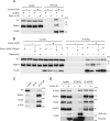
Mammalian Tti1 interacts with mTOR. A, HEK293T cells were transfected with an empty vector or FLAG-mTOR. Two days later, cells were harvested and lysed in a buffer containing either 0.3% CHAPS or 0.5% Triton X-100. Immunoprecipitation was then performed with anti-FLAG antibody. The lysate lanes contain one-twelfth of the lysate volume used for immunoprecipitation. B, HEK293T cells were transfected with an empty vector or FLAG-mTOR. Two days later, cells were cultured in an amino acid-free DMEM (without FBS) for 120 min and then cultured in a complete medium for 30 min. Rapamycin treatment was performed by incubation for 30 min with a complete medium containing 20 μg/ml rapamycin. Cells were lysed, and immunoprecipitation was performed with anti-FLAG antibody. The lysate lanes contain one-twelfth of the lysate volume used for immunoprecipitation. C, HeLa cells were lysed, and immunoprecipitation was performed with the indicated antibodies to detect endogenous interactions. The lysate lanes contain one-four-hundredth of the lysate volume used for immunoprecipitation. D, HEK293T cells were transfected with an empty vector or FLAG-Tti1. Two days later, cells were lysed, and immunoprecipitation was performed with anti-FLAG and anti-mTOR antibodies. The lysate lanes contain one-sixth or one-sixtieth of the lysate volume used for immunoprecipitation with anti-FLAG or anti-mTOR antibody, respectively.
FIGURE 3.
FIGURE 3.
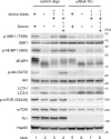
Tti1 positively regulates mTOR activity. HeLa cells transfected with Tti1 siRNA or control siRNA were cultured in amino acid-free DMEM (without FBS) for 120 min, and then cultured in regular DMEM with or without 10% FBS for 30 min.
FIGURE 4.
FIGURE 4.
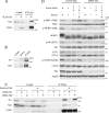
Tti1 interacts with Tel2 and is important for Tel2–mTOR association. A, HEK293T cells were transfected with an empty vector or FLAG-Tti1. Two days later, cells were lysed and immunoprecipitation was performed with anti-FLAG antibody. The lysate lanes contain one-twelfth of the lysate volume used for immunoprecipitation. B, endogenous interaction between Tti1 and Tel2 was determined in HeLa cells. Immunoprecipitation was performed with anti-Tel2 antiserum or preimmune rabbit serum (Pre). The lysate lanes contain one-twentieth of the lysate volume used for immunoprecipitation. C, HEK293T cells were transfected with siRNA against Tti1 or Tel2, and then with an empty vector or FLAG-mTOR. Cells were lysed and immunoprecipitation was performed with anti-FLAG antibody. The lysate lanes contain one-fifteenth of the lysate volume used for immunoprecipitation. D, HeLa cells transfected with Tel2 siRNA or control siRNA were treated as in Fig. 2.
FIGURE 5.
FIGURE 5.
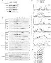
Tti1 and Tel2 interact with and stabilize PIKKs. A, HEK293T cells were transfected with empty vector, FLAG-Tti1, or FLAG-Tel2. Two days later, cells were lysed, and immunoprecipitation was performed with anti-FLAG antibody. The lysate lanes contain one-twenty fourth of the lysate volume used for immunoprecipitation. B, HeLa cells were treated with siRNAs against Tti1 or/and Tel2 for 4 or 6 days. Transfection was performed every other day. Cells were then harvested, and immunoblot analysis was performed with the indicated antibodies.
FIGURE 6.
FIGURE 6.
Tti1 and Tel2 are important for mTOR complex assembly. A, HeLa cells transfected with Tti1 or control siRNAs were lysed, and immunoprecipitation was performed with the indicated antibodies. The lysate lanes contain one-twentieth of the lysate volume used for immunoprecipitation. B–D, HeLa cells transfected with the indicated siRNAs or treated with 0.8% 1-butanol for 3 h were lysed. The cytosolic fractions of the cells were subjected to gel filtration analysis using a Superose 6 column (B) or subjected to Western blot analysis (D). Immunoblot signals in B were quantified with ImageJ and plotted (C). The value of the total signal was adjusted to the amount of total protein of non-treated samples for each protein, as estimated from Western blot analysis of total cell lysates (D).

References

    1. Bakkenist C. J., Kastan M. B. (2004) Cell 118, 9–17 - PubMed
    1. Lempiäinen H., Halazonetis T. D. (2009) EMBO J. 28, 3067–3073 - PMC - PubMed
    1. Lovejoy C. A., Cortez D. (2009) DNA Repair 8, 1004–1008 - PMC - PubMed
    1. Wullschleger S., Loewith R., Hall M. N. (2006) Cell 124, 471–484 - PubMed
    1. Guertin D. A., Sabatini D. M. (2007) Cancer Cell 12, 9–22 - PubMed

Publication types

MeSH terms