. 2010 Jul;84(13):6711-9.
doi: 10.1128/JVI.00547-10. Epub 2010 Apr 28.

Rearranged genomic RNA segments offer a new approach to the reverse genetics of rotaviruses

Affiliations

Rearranged genomic RNA segments offer a new approach to the reverse genetics of rotaviruses

Cécile Troupin et al. J Virol. 2010 Jul.

Abstract

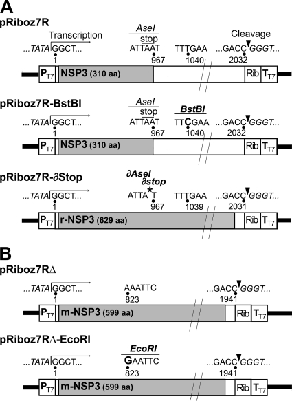
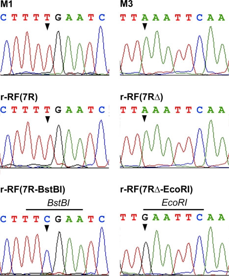
Group A rotaviruses (RV), members of the Reoviridae family, are a major cause of infantile acute gastroenteritis. The RV genome consists of 11 double-stranded RNA segments. In some cases, an RNA segment is replaced by a rearranged RNA segment, which is derived from its standard counterpart by partial sequence duplication. We report here a reverse genetics system for RV based on the preferential packaging of rearranged RNA segments. Using this system, wild-type or in vitro-engineered forms of rearranged segment 7 from a human rotavirus (encoding the NSP3 protein), derived from cloned cDNAs and transcribed in the cytoplasm of COS-7 cells with the help of T7 RNA polymerase, replaced the wild-type segment 7 of a bovine helper virus (strain RF). Recombinant RF viruses (i.e., engineered monoreassortant RF viruses) containing an exogenous rearranged RNA were recovered by propagating the viral progeny in MA-104 cells, with no need for additional selective pressure. Our findings offer the possibility to extend RV reverse genetics to segments encoding nonstructural or structural proteins for which no potent selective tools, such as neutralizing antibodies, are available. In addition, the system described here is the first to enable the introduction of a mutated gene expressing a modified nonstructural protein into an infectious RV. This reverse genetics system offers new perspectives for investigating RV protein functions and developing recombinant live RV vaccines containing specific changes targeted for attenuation.


Figures

FIG. 1.
FIG. 1.
pRiboz plasmids constructs containing 7R (A) and 7RΔ (B) derivatives. All pRiboz constructs contain a full-length rearranged segment 7 cDNA flanked by the T7pol promoter (PT7) and by the HDV ribozyme (Rib), followed by the T7pol terminator (TT7). Numbers indicate the nucleotide position in the rearranged segment, open boxes indicate the 5′- and 3′-UTRs, and gray boxes indicate the NSP3 coding sequence. Arrows and arrowheads indicate the transcription initiation site and the ribozyme self-cleavage site, respectively. The nucleotide changes used to create BstBI and EcoRI sites are indicated in bold. An asterisk indicates the A967 deletion that abrogates the stop codon and AseI site in 7R-∂Stop.
FIG. 2.
FIG. 2.
Rescue of recombinant RF viruses containing cDNA-derived rearranged segment 7R. Viral progenies produced in COS-7 cells transfected with pRiboz7R plasmid were serially propagated in MA-104 cells, and viral dsRNA was analyzed at each passage, by RT-PCR using primers DPZJ3 and RPZJ3 (A) and by PAGE (B). Pn indicates the passage number. Recombinant virus r-RF(7R) is a representative clone rescued from MA-104 cell passage 18. dsRNAs prepared from M1 and RF viruses were used as controls. Arrows indicate the location of segment 7R in M1 and recombinant RF viruses. Mw, 100-bp-ladder molecular size marker.
FIG. 3.
FIG. 3.
Rescue of recombinant RF viruses containing cDNA-derived rearranged segment 7RΔ. Viral progenies produced in COS-7 cells transfected by pRiboz7RΔ plasmid were serially propagated in MA-104 cells, and viral dsRNA was analyzed at each passage, by RT-PCR using primers DPZJ3 and RPZJ3 (A) and by PAGE (B). Pn indicates the passage number. Recombinant virus r-RF(7RΔ) is a representative clone rescued from MA-104 cell passage 18. dsRNAs prepared from M3 and RF viruses were used as controls. Arrows indicate the location of segment 7RΔ in M3 and recombinant RF viruses. Mw, 100-bp-ladder molecular size marker.
FIG. 4.
FIG. 4.
Rescue of recombinant RF viruses containing in vitro-modified, cDNA-derived rearranged segment 7R or 7RΔ. Recombinant viruses r-RF(7R), r-RF(7R-BstBI), r-RF(7RΔ), and r-RF(7RΔ-EcoRI) are representative clones rescued from MA-104 cell culture. (A) dsRNA profiles of recombinant and control (M1 [7R], M3 [7RΔ], and RF) viruses. Segments 7R and 7RΔ are indicated by arrows. (B) BstBI and EcoRI restriction profiles of RT-PCR products obtained from segments 7 of recombinant and control viruses. +, digested; −, not digested. Numbers indicate the expected sizes of fragments. Mw, 100-bp-ladder molecular size marker.
FIG. 5.
FIG. 5.
Site-specific mutations in segments 7 of recombinant RF viruses. Sequence electropherograms are shown for RT-PCR products obtained from recombinant viruses r-RF(7R), r-RF(7R-BstBI), r-RF(7RΔ), and r-RF(7RΔ-EcoRI) and from control viruses M1 (7R) and M3 (7RΔ). Arrowheads indicate nucleotides changes that created BstBI and EcoRI sites in r-RF(7R-BstBI) and r-RF(7RΔ-EcoRI), respectively.
FIG. 6.
FIG. 6.
Rescue of a recombinant RF virus containing an in vitro-modified, cDNA-derived rearranged segment 7R encoding a duplicated NSP3 protein. (A) dsRNA profiles of a representative r-RF(7R-∂Stop) viral clone rescued from MA-104 cell passage 18 and of control viruses RF and M1 (7R). An arrow indicates the location of segment 7R. (B) Electrophoretic analysis of RT-PCR products obtained from r-RF(7R-∂Stop) and M1 viruses, digested (+) or not (−) by AseI (numbers indicate the expected sizes of fragments). Mw, 100-bp-ladder molecular size marker. (C) Sequence electropherograms for RT-PCR products obtained from M1 (7R) and r-RF(7R-∂Stop) viruses. The arrowhead indicates the A967 residue deleted in the AseI site and the NSP3 stop codon. The predicted aa translation is indicated above the nucleotide sequences. For 7R-∂Stop (encoding r-NSP3), the 9 aa linking the last aa (YD) of NSP3 to the first aa (ME) of the duplicated NSP3 sequence are boxed. (D) Western blot analysis of r-RF(7R-∂Stop) and RF virus-infected cell lysates, using the ID3 monoclonal antibody specific for NSP3. Arrows indicate the NSP3 and r-NSP3 proteins, expressed by the RF and r-RF(7R-∂Stop) viruses, respectively. Numbers indicate molecular size, in kilodaltons.

References

    1. Aponte, C., N. M. Mattion, M. K. Estes, A. Charpilienne, and J. Cohen. 1993. Expression of two bovine rotavirus non-structural proteins (NSP2, NSP3) in the baculovirus system and production of monoclonal antibodies directed against the expressed proteins. Arch. Virol. 133:85-95. - PubMed
    1. Boyce, M., C. C. Celma, and P. Roy. 2008. Development of reverse genetics systems for bluetongue virus: recovery of infectious virus from synthetic RNA transcripts. J. Virol. 82:8339-8348. - PMC - PubMed
    1. Chizhikov, V., and J. T. Patton. 2000. A four-nucleotide translation enhancer in the 3′-terminal consensus sequence of the nonpolyadenylated mRNAs of rotavirus. RNA 6:814-825. - PMC - PubMed
    1. Dehee, A., N. Schnepf, C. Deback, A. L. Poeury, and A. Garbarg-Chenon. 2006. Preferential segregation of rearranged segments in rotaviral progeny after mixed infection, abstr. PW4.1, p. 81. Abstr. 9th Int. Symp. ds-RNA Viruses.
    1. Desselberger, U. 1996. Genome rearrangements of rotaviruses. Adv. Virus Res. 46:69-95. - PubMed

Publication types

LinkOut - more resources